Copyright
©The Author(s) 2020.
World J Gastrointest Oncol. Jul 15, 2020; 12(7): 768-781
Published online Jul 15, 2020. doi: 10.4251/wjgo.v12.i7.768
Published online Jul 15, 2020. doi: 10.4251/wjgo.v12.i7.768
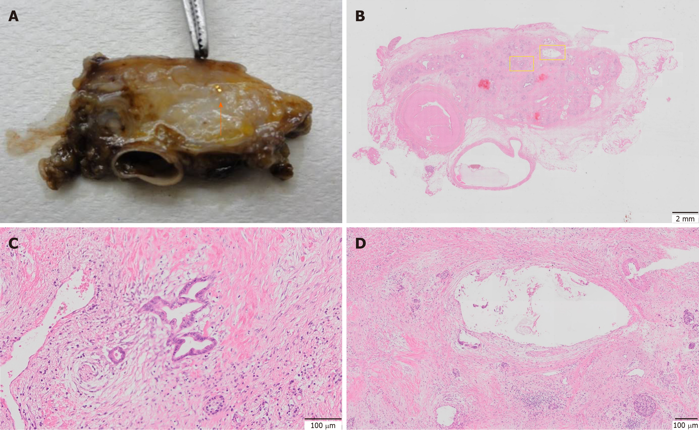
Figure 6 Histological findings.
A: Orange arrow indicates the gold marker; B: hematoxylin and eosin staining of the pathological specimen after chemoradiation therapy. Left yellow square indicates magnified area shown in C and right yellow square indicates magnified area shown in D; C: Very few residual cancer observed with Evans grade III status; D: The hollow area is where the marker was inserted. No significant changes around this area, such as accumulation of inflammatory cells or fibrosis, were observed.
- Citation: Ashida R, Fukutake N, Takada R, Ioka T, Ohkawa K, Katayama K, Akita H, Takahashi H, Ohira S, Teshima T. Endoscopic ultrasound-guided fiducial marker placement for neoadjuvant chemoradiation therapy for resectable pancreatic cancer. World J Gastrointest Oncol 2020; 12(7): 768-781
- URL: https://www.wjgnet.com/1948-5204/full/v12/i7/768.htm
- DOI: https://dx.doi.org/10.4251/wjgo.v12.i7.768


