Copyright
©The Author(s) 2020.
World J Gastrointest Oncol. Apr 15, 2020; 12(4): 457-466
Published online Apr 15, 2020. doi: 10.4251/wjgo.v12.i4.457
Published online Apr 15, 2020. doi: 10.4251/wjgo.v12.i4.457
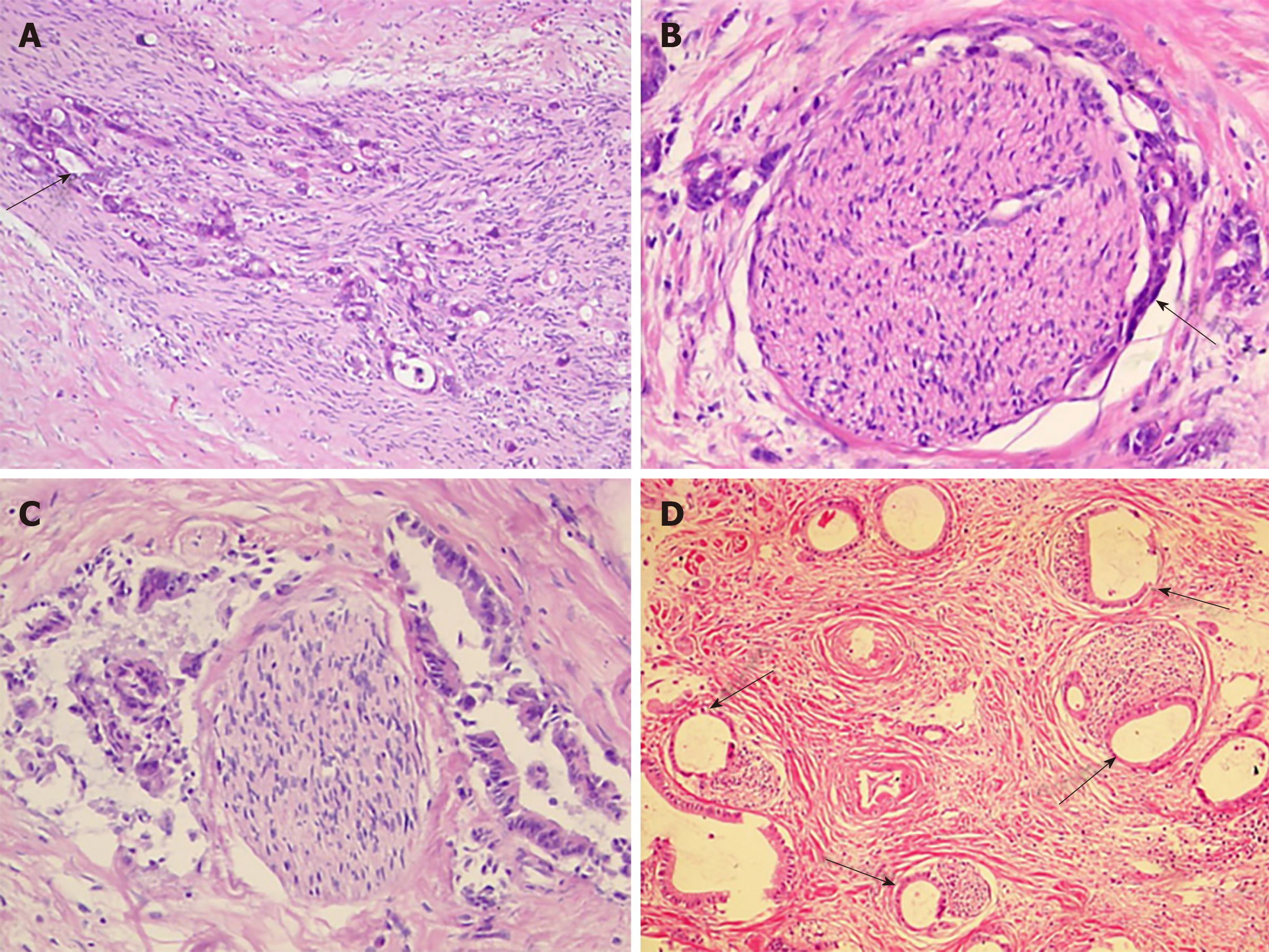
Figure 1 Perineural invasion in human hilar cholangiocarcinoma.
A: Tumor cells (arrow) located within the peripheral nerve sheath in clusters [Hematoxylin-eosin (HE) staining, ×100]; B: Tumor cells (arrow) from glandular elements in perineural space (HE staining, ×100); C: Tumor cells invaded more than 33% of the circumference of the nerve (HE staining, ×100); D: Modes of perineural invasion (arrows) in one patient (HE staining, ×100).
- Citation: Li CG, Zhou ZP, Tan XL, Zhao ZM. Perineural invasion of hilar cholangiocarcinoma in Chinese population: One center’s experience. World J Gastrointest Oncol 2020; 12(4): 457-466
- URL: https://www.wjgnet.com/1948-5204/full/v12/i4/457.htm
- DOI: https://dx.doi.org/10.4251/wjgo.v12.i4.457


