Copyright
©The Author(s) 2019.
World J Gastrointest Oncol. Aug 15, 2019; 11(8): 599-621
Published online Aug 15, 2019. doi: 10.4251/wjgo.v11.i8.599
Published online Aug 15, 2019. doi: 10.4251/wjgo.v11.i8.599
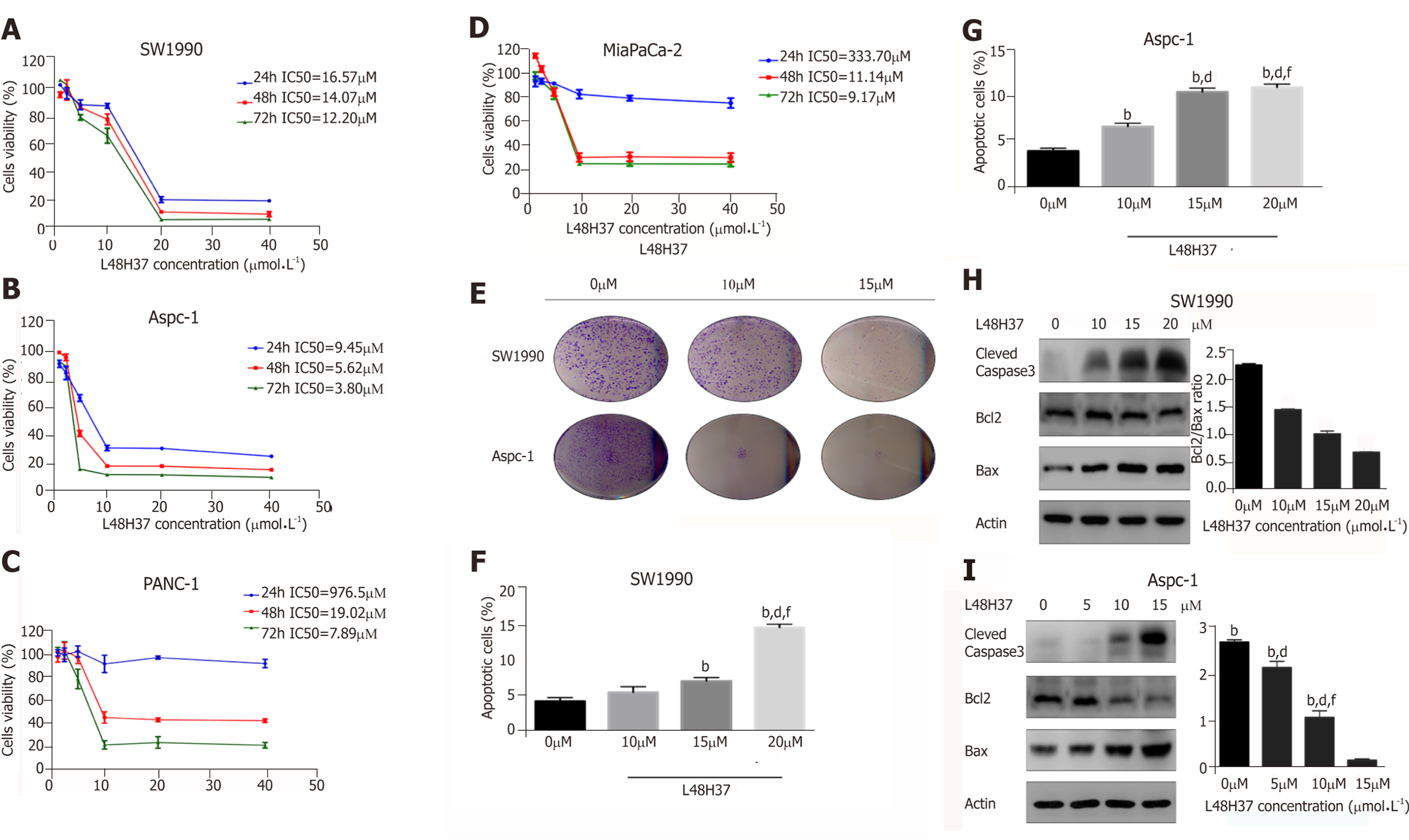
Figure 1 L48H37 inhibits proliferation and promotes apoptosis in pancreatic cancer cells.
A-D: Percentage of viable SW1990, ASPC-1, PANC-1 and MIA PaCa2 cells incubated with different concentrations of L48H37 (1.25, 2.5, 5, 10, 20 and 40 μmol/L) or DMSO (negative control) for 24, 48 and 72 h. The IC50 values in the different cell lines are shown; E: Representative pictures of colonies from SW1990 and ASPC-1 cells treated with increasing concentrations of L48H37 for 24 h; F-G: SW1990 and Aspc-1 cells were treated with various concentrations of L48H37 for 24 h. Cell apoptosis was detected by flow cytometry. Histogram illustrating the rate of apoptosis cells. Data were expressed as mean ± SEM; F: bP < 0.01 vs L48H37 (0 μmol/L) group. dP < 0.01 vs L48H37 (10 μmol/L) group. fP < 0.01 vs L48H37 (15 μmol/L) group; G: bP < 0.01 vs L48H37 (0 μmol/L) group; dP < 0.01 vs L48H37 (5 μmol/L) group. fP < 0.01 vs L48H37 (10 μmol/L) group; H-I: Western blotting showing expression levels of Cleaved caspase3, Bcl-2 and Bax proteins in SW1990 and Aspc-1 cells following treatment with DMSO or L48H37 for 12 h. Grayscale values of Bcl-2 and Bax were measured relative to β-actin, and the ratio of Bcl-2/Bax expression was calculated. Data were expressed as mean ± SEM; H: bP < 0.01 vs L48H37 (0 μmol/L) group. dP < 0.01 vs L48H37 (10 μmol/L) group. fP < 0.01 vs L48H37 (15 μmol/L) group; I: bP < 0.01 vs L48H37 (0 μmol/L) group. dP < 0.01 vs L48H37 (5 μmol/L) group. fP < 0.01 vs L48H37 (10 μmol/L) group.
- Citation: Li SS, Jiang WL, Xiao WQ, Li K, Zhang YF, Guo XY, Dai YQ, Zhao QY, Jiang MJ, Lu ZJ, Wan R. KMT2D deficiency enhances the anti-cancer activity of L48H37 in pancreatic ductal adenocarcinoma. World J Gastrointest Oncol 2019; 11(8): 599-621
- URL: https://www.wjgnet.com/1948-5204/full/v11/i8/599.htm
- DOI: https://dx.doi.org/10.4251/wjgo.v11.i8.599


