Copyright
©The Author(s) 2019.
World J Gastrointest Oncol. Nov 15, 2019; 11(11): 971-982
Published online Nov 15, 2019. doi: 10.4251/wjgo.v11.i11.971
Published online Nov 15, 2019. doi: 10.4251/wjgo.v11.i11.971
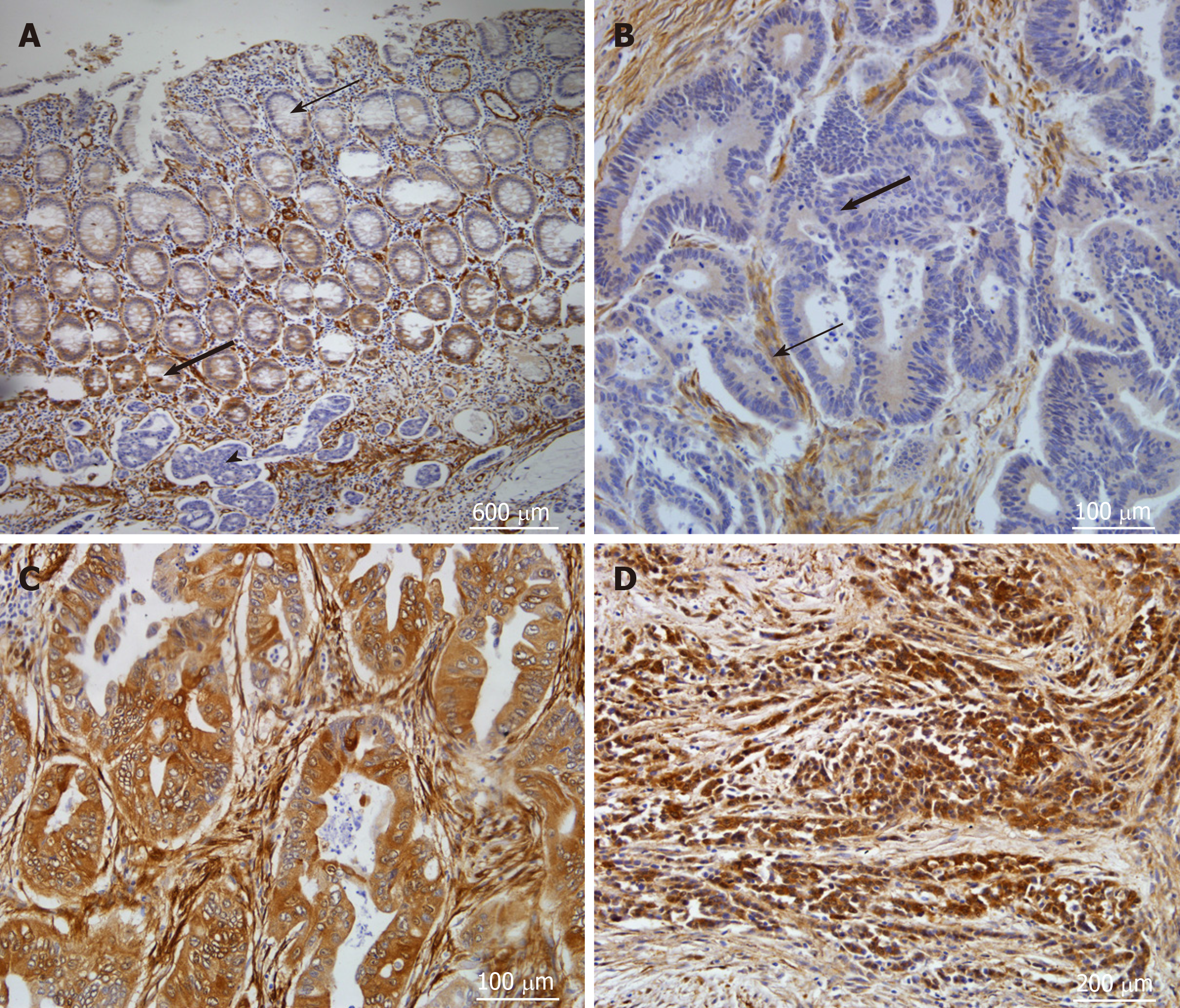
Figure 4 CNN3 expression in formalin-fixed, paraffin-embedded tissues from resected colorectal cancers.
A: The normal mucosa showed positive expression at the crypt bases (thick arrow) and faint to negative expression superficially (thin arrow, Magnification 40×). Note the negative tumor islets below the mucosa (arrow head); B: Well differentiated colon adenocarcinoma showing negative expression of CNN3 (thick arrow) while the stroma showed positive expression (thin arrow) serving as internal positive control (Magnification 200×); C: Well differentiated colon adenocarcinoma showing positive CNN3 expression (Magnification 200×); D: Poorly differentiated rectal adenocarcinoma showing intense positive expression (Magnification 100×). Chromogen, DAB marks the positive expression with brown color; counterstain, Mayer’s hematoxylin. CNN3: Calponin 3.
- Citation: Nair VA, Al-khayyal NA, Sivaperumal S, Abdel-Rahman WM. Calponin 3 promotes invasion and drug resistance of colon cancer cells. World J Gastrointest Oncol 2019; 11(11): 971-982
- URL: https://www.wjgnet.com/1948-5204/full/v11/i11/971.htm
- DOI: https://dx.doi.org/10.4251/wjgo.v11.i11.971


