Copyright
©2010 Baishideng Publishing Group Co.
World J Gastrointest Oncol. Dec 15, 2010; 2(12): 443-445
Published online Dec 15, 2010. doi: 10.4251/wjgo.v2.i12.443
Published online Dec 15, 2010. doi: 10.4251/wjgo.v2.i12.443
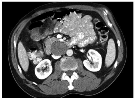
Figure 1 Computed tomography image revealing a large mass in the retroperitoneum abutting the pancreas and compressing the inferior vena cava.
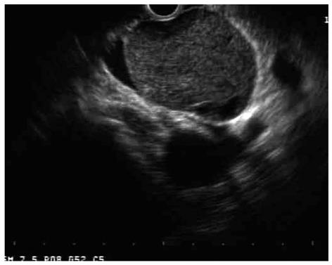
Figure 2 Endoscopic ultrasound image of a well-circumscribed hypoechoic mass compressing the inferior vena cava and aorta.
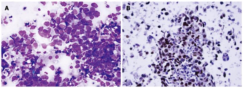
Figure 3 Photomicrographs.
A: A fine needle aspirate revealing undifferentiated anaplastic carcinoma of unclear origin (DiffQuik™ stain, × 400); B: A fine needle aspirate revealing a strong nuclear immunostaining pattern consistent with a germ cell tumor, favoring seminoma (OCT-4 stain - HE counterstain, × 200).
- Citation: Womeldorph CM, Zalupski MM, Knoepp SM, Soltani M, Elmunzer BJ. Retroperitoneal germ cell tumor diagnosed by endoscopic ultrasound-guided fine needle aspiration. World J Gastrointest Oncol 2010; 2(12): 443-445
- URL: https://www.wjgnet.com/1948-5204/full/v2/i12/443.htm
- DOI: https://dx.doi.org/10.4251/wjgo.v2.i12.443


