Copyright
©The Author(s) 2024.
World J Gastrointest Oncol. Mar 15, 2024; 16(3): 773-786
Published online Mar 15, 2024. doi: 10.4251/wjgo.v16.i3.773
Published online Mar 15, 2024. doi: 10.4251/wjgo.v16.i3.773
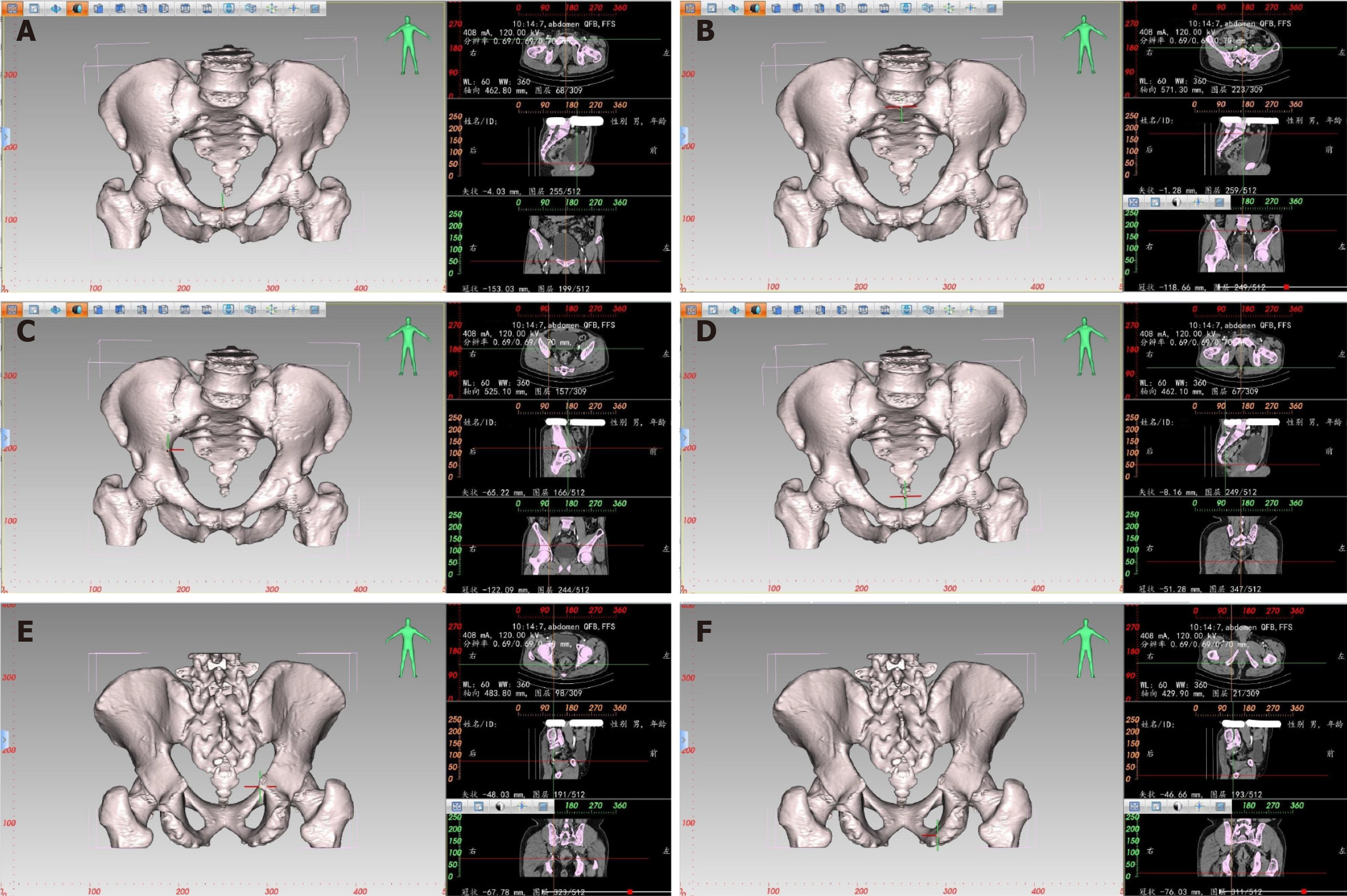
Figure 1 Localization diagram of each positioning point during three-dimensional pelvic measurement.
A: Localization diagram of the highest point of the pubic symphysis during three-dimensional (3D) pelvic measurement (left); positioning of the corresponding measurement point at the intersection of two axis lines on the transverse, sagittal, and coronal planes (right); B: Localization diagram of the midpoint of the anterior edge of the sacral promontory during 3D pelvic measurement (left); positioning of the corresponding measurement point at the intersection of two axis lines on the transverse, sagittal, and coronal planes (right); C: Localization diagram of the maximum distance between the left and right iliopectineal line during 3D pelvic measurement (left); positioning of the corresponding measurement point at the intersection of two axis lines on the transverse, sagittal, and coronal planes (right); D: Localization diagram of the tip of the coccyx during 3D pelvic measurement (left); positioning of the corresponding measurement point at the intersection of two axis lines on the transverse, sagittal, and coronal planes (right); E: Localization diagram of the ischial spine during 3D pelvic measurement (left); positioning of the corresponding measurement point at the intersection of two axis lines on the transverse, sagittal, and coronal planes (right); F: Localization diagram of the ischial tuberosity during 3D pelvic measurement (left); positioning of the corresponding measurement point at the intersection of two axis lines on the transverse, sagittal, and coronal planes (right).
Figure 2 Illustration of three-dimensional pelvic reconstruction in a male patient.
A: Mid-sagittal lateral view: (AB) anterior-posterior diameter of pelvic inlet, (CD) anterior-posterior diameter of mid-pelvis, (CE) anterior-posterior diameter of pelvic outlet, (AC) superior-inferior diameter of the pubic symphysis, (BE) sacrococcygeal distance, (BD) superior-inferior diameter of sacrum, (AE) superior pubococcygeal diameter, (FG) anterior-posterior sacropubic distance, (FI) sacrococcygeal curvature height, (FH) sacral curvature height, (α) sacrococcygeal angle, (β) sacropubic angle; B: Anteroposterior view: (PQ) transverse diameter of pelvic inlet and (LM) interischial spine diameter; C: Posteroanterior view: (LM) interischial spine diameter and (NO) interischial tuberosity diameter.
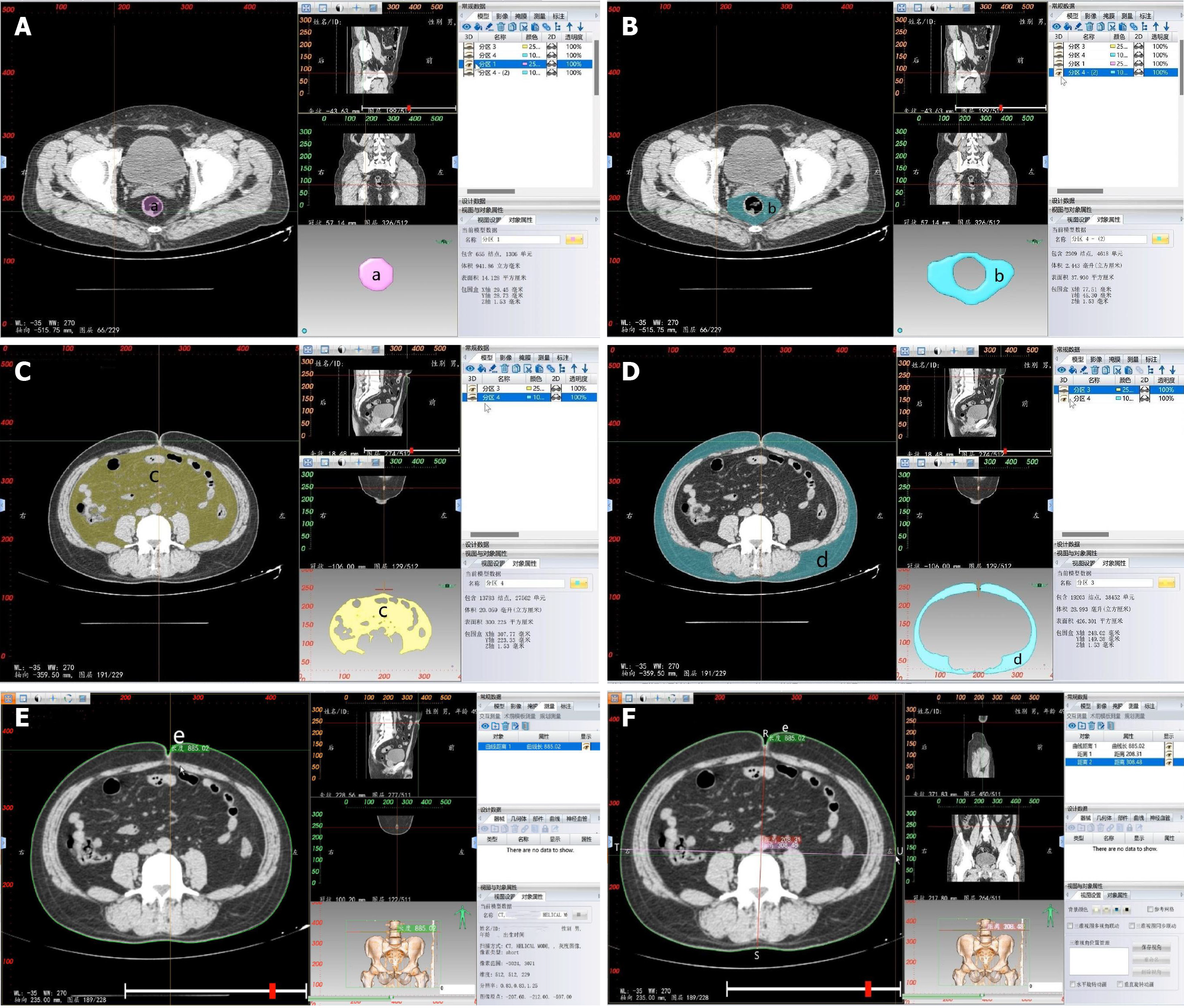
Figure 3 Measurements of soft tissue parameters.
A: Measurement of rectal area (a): Rectal area at the ischial spine level; B: Measurement of mesorectal fat area (b): Mesorectal fat area at the ischial spine level; C: Measurement of visceral fat area (c): Visceral fat area at the level of the umbilicus; D: Measurement of subcutaneous fat area (d): Subcutaneous fat area at the level of the umbilicus; E: Measurement of waist circumference (e): Waist circumference at the level of the umbilicus; F: Measurement of anterior-posterior abdominal diameter (RS): It refers to the distance measured from the line connecting the umbilicus to the spinous process at the level of the umbilicus, between the abdominal wall section and the lumbar section; measurement of transverse abdominal diameter (TU): It refers to the distance measured from the level of the umbilicus to the horizontal line at the anterior edge of the lumbar vertebrae.
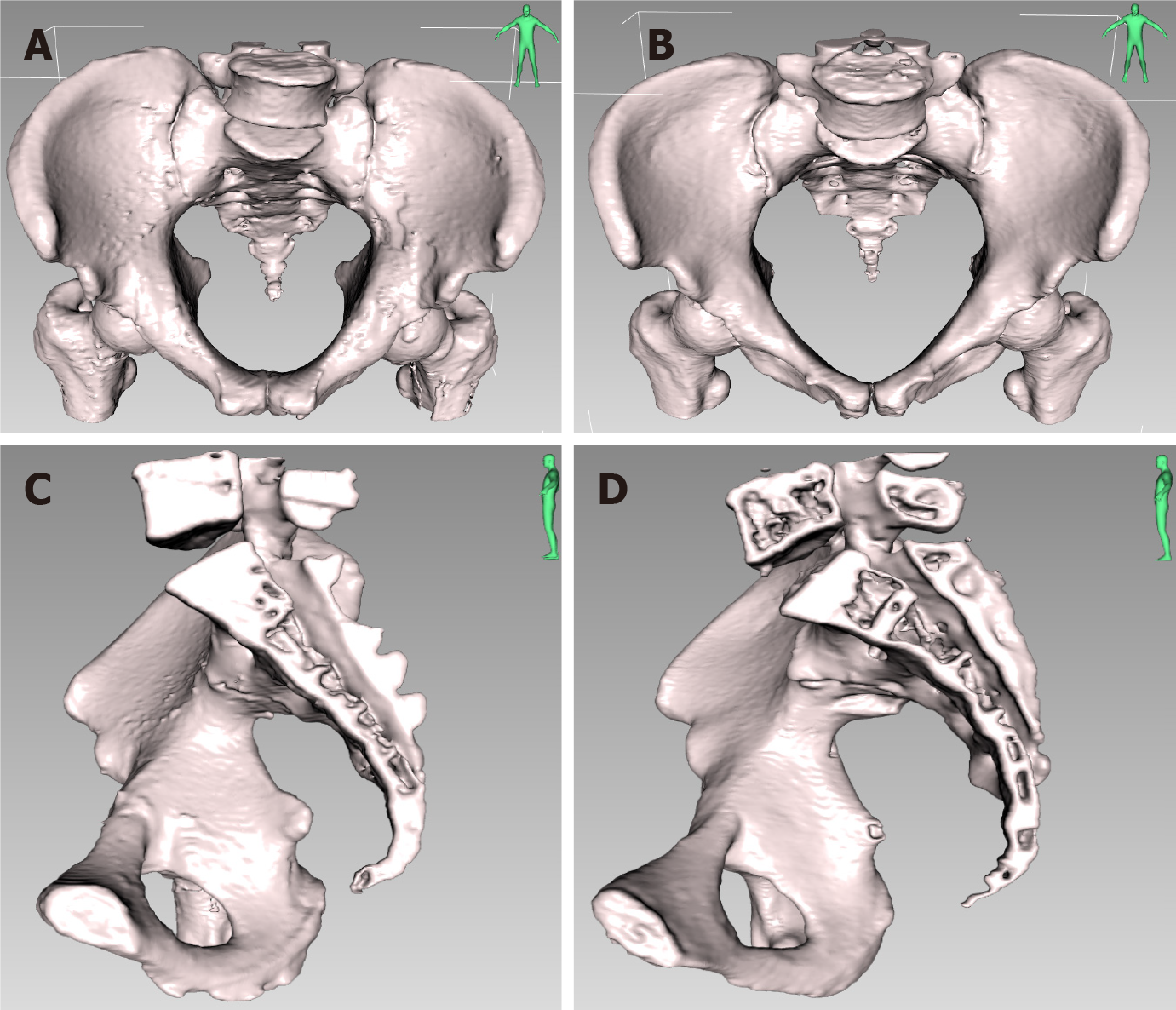
Figure 4 Front and lateral view of the three-dimensional pelvic reconstruction between male and female patients in the forward tilt position and the mid-sagittal position respectively.
A: Front view of the three-dimensional (3D) pelvic reconstruction in a male patient in the forward tilt position; B: Front view of the 3D pelvic reconstruction in a female patient in the forward tilt position; C: Lateral view of the 3D pelvic reconstruction in a male patient in the mid-sagittal position; D: Lateral view of the 3D pelvic reconstruction in a female patient in the mid-sagittal position. The male pelvis is deep and narrow, with a forward tilt, straighter sacrum, and a higher overall curvature. The female pelvis is wide and shallow, with a backward tilt and a smaller overall curvature.
- Citation: Zhou XC, Ke FY, Dhamija G, Chen H, Wang Q. Study on sex differences and potential clinical value of three-dimensional computerized tomography pelvimetry in rectal cancer patients. World J Gastrointest Oncol 2024; 16(3): 773-786
- URL: https://www.wjgnet.com/1948-5204/full/v16/i3/773.htm
- DOI: https://dx.doi.org/10.4251/wjgo.v16.i3.773


