Copyright
©The Author(s) 2024.
World J Gastrointest Oncol. Mar 15, 2024; 16(3): 1084-1090
Published online Mar 15, 2024. doi: 10.4251/wjgo.v16.i3.1084
Published online Mar 15, 2024. doi: 10.4251/wjgo.v16.i3.1084
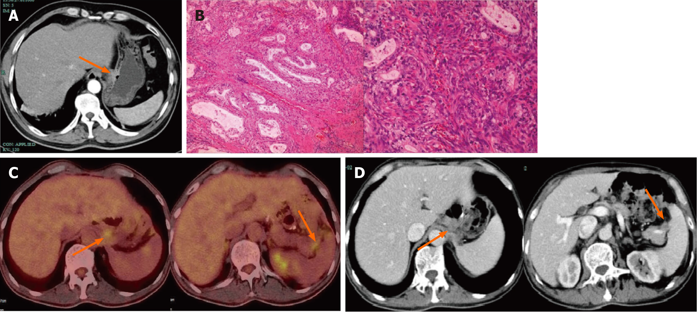
Figure 1 Computed tomography scans taken before and after treatment and Pathology of surgical specimens after total gastrectomy.
A: Enhanced computed tomography (CT) scan of the abdomen before surgery: Thickening of the gastric wall near the lesser curvature of the stomach and formation of ulcers; B: Pathology of surgical specimens after total gastrectomy: Histological type: moderately differentiated adenocarcinoma; Lauren’s classification: Intestinal type; Infiltration depth: Infiltration to the sub-serosal layer, involving nerves; C: Abdominal lymph node metastasis positron emission tomography-CT: Enlarged lymph nodes in the left upper peritoneum and splenic and pancreatic space; D: Enhanced CT scan of the abdomen after 6 cycles of treatment: Lymph node reduction compared to before treatment. The orange arrows showed the location and range of the tumor.
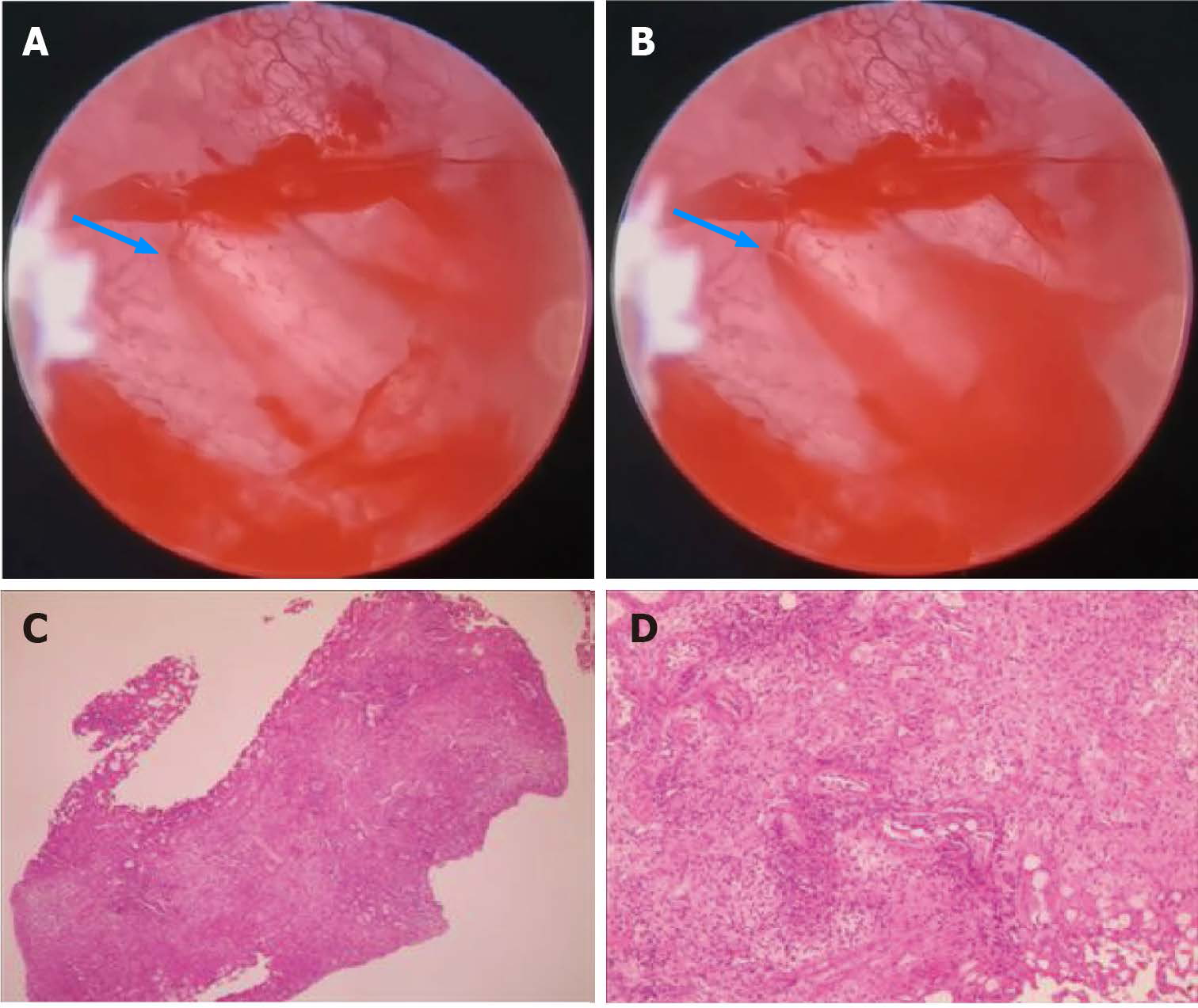
Figure 2 Cystoscopy and pathological results of biopsy under cystoscopy.
A and B: Cystoscopy shows bleeding of the bladder mucosa (blue arrow); C and D: Pathological results of biopsy under cystoscopy showed that the lesion was tumor-free.
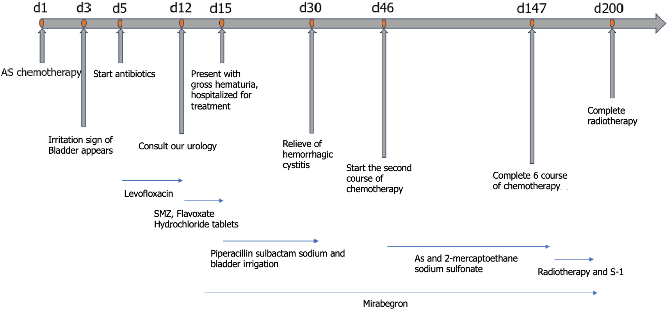
Figure 3 Timeline of chemotherapy, urologic symptoms, and concomitant treatment.
SMZ: Sulfamethoxazole; AS: Nab-paclitaxel combined with S-1.
- Citation: Zhang XJ, Lou J. Hemorrhagic cystitis in gastric cancer after nanoparticle albumin-bound paclitaxel: A case report. World J Gastrointest Oncol 2024; 16(3): 1084-1090
- URL: https://www.wjgnet.com/1948-5204/full/v16/i3/1084.htm
- DOI: https://dx.doi.org/10.4251/wjgo.v16.i3.1084


