Copyright
©The Author(s) 2020.
World J Gastrointest Oncol. Jan 15, 2020; 12(1): 92-100
Published online Jan 15, 2020. doi: 10.4251/wjgo.v12.i1.92
Published online Jan 15, 2020. doi: 10.4251/wjgo.v12.i1.92
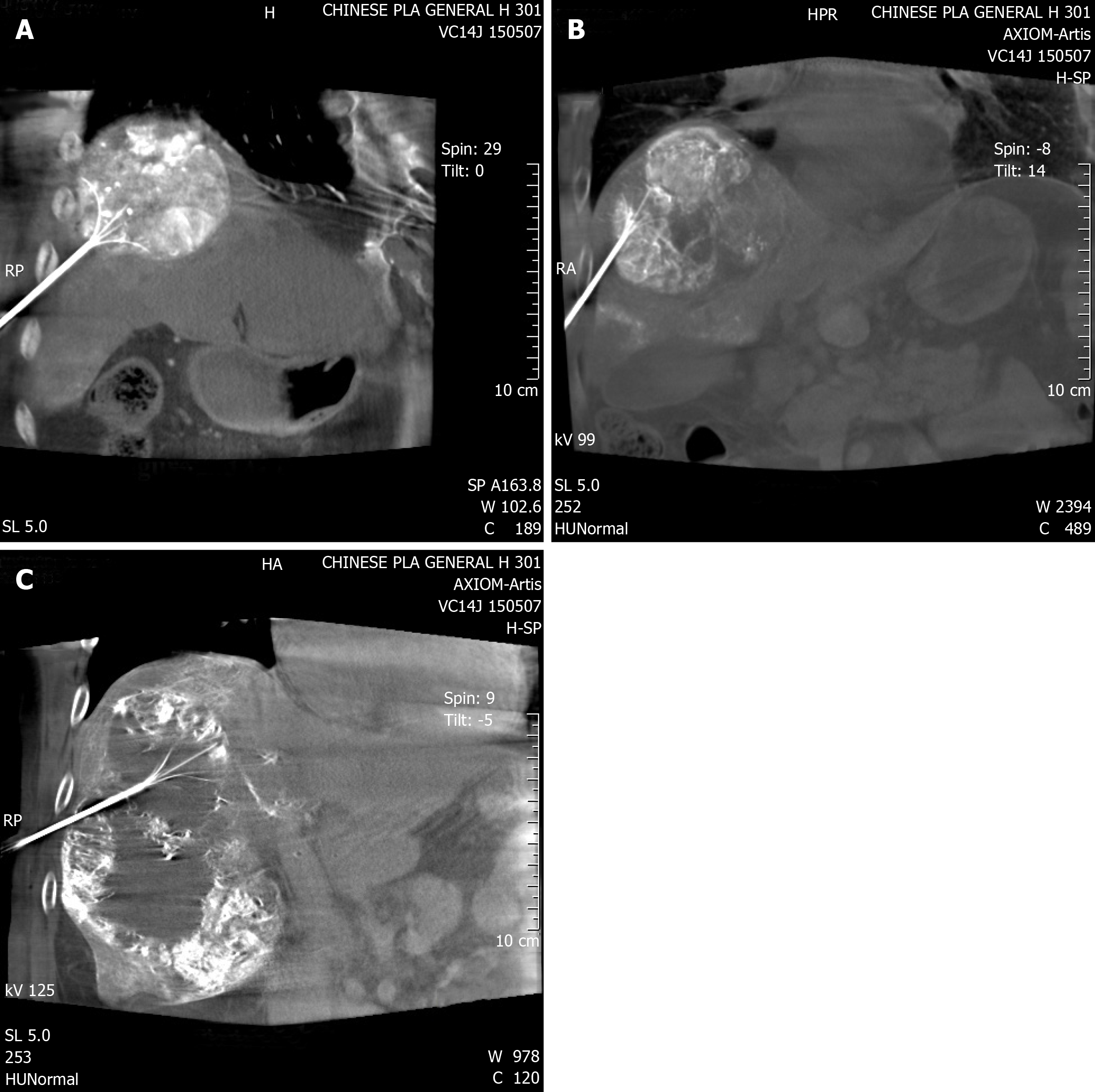
Figure 1 Cone-beam computed tomography image confirmed the position of the radiofrequency probe.
A-C: Radiofrequency probe inserted at an angle to avoid lung damage.
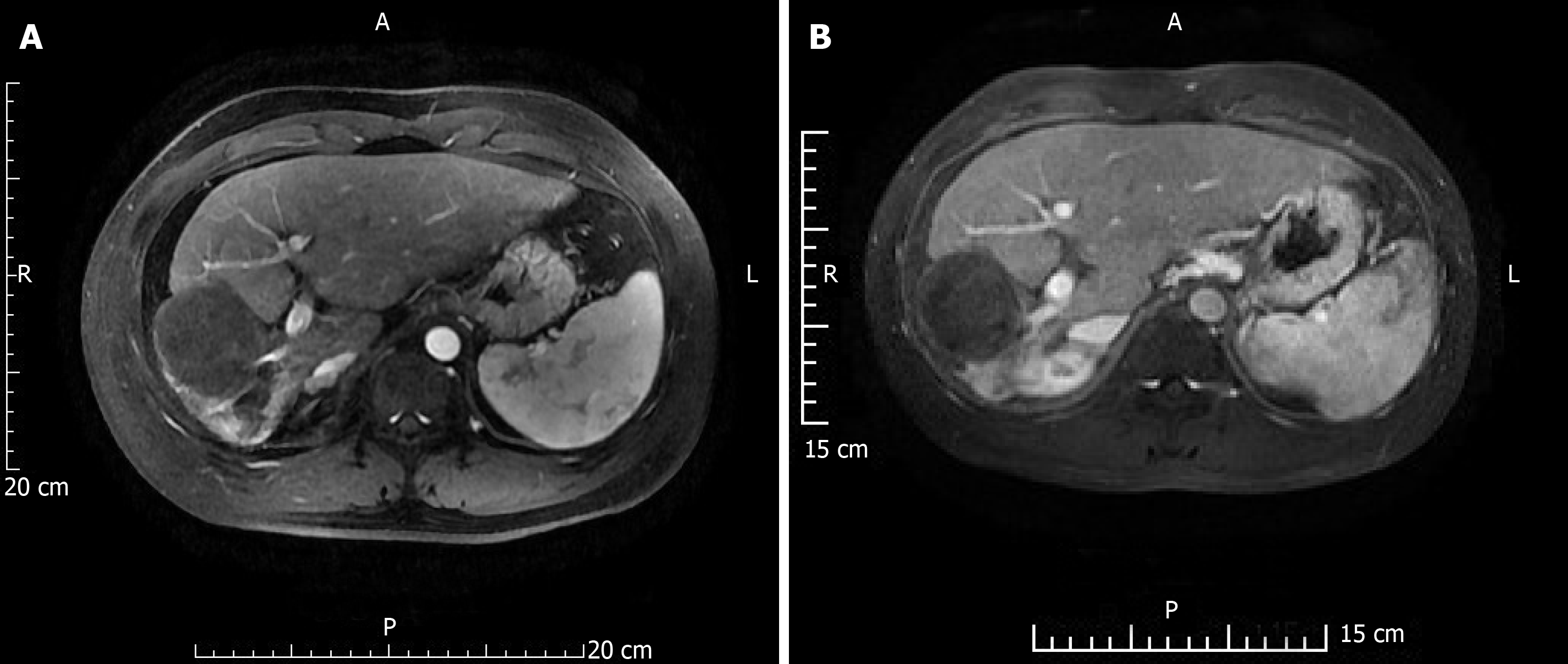
Figure 2 A 41-year-old male patient re-examined 2 and 4 years after combination therapy.
A: Magnetic resonance imaging (MRI) at 2 years; B: MRI at 4 years. Hepatocellular carcinoma lesions showed pyknosis and necrosis.
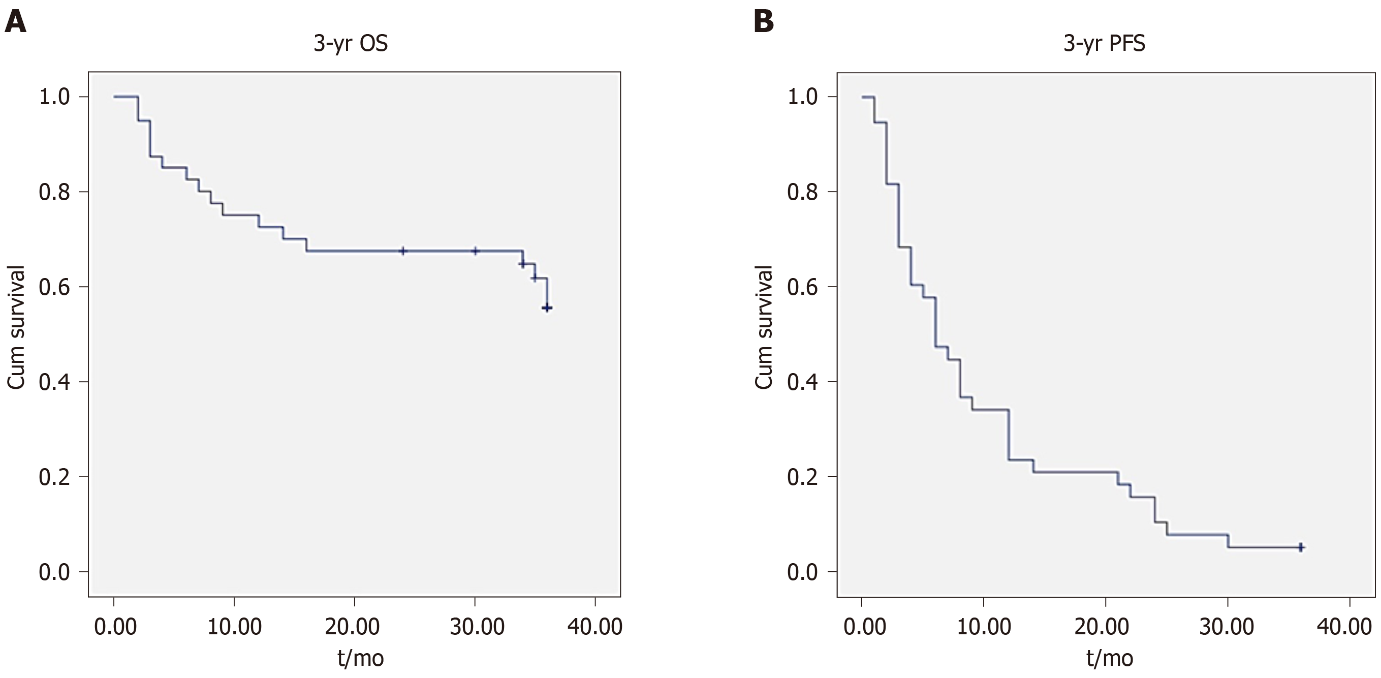
Figure 3 Kaplan–Meier analysis of overall survival and progression-free survival.
Kaplan–Meier survival curves shown for patients with large hepatocellular carcinomas treated with combination therapy. A: 3-year overall survival; B: 3-year progression-free survival. OS: Overall survival; PFS: Progression-free survival.
- Citation: Duan F, Bai YH, Cui L, Li XH, Yan JY, Wang MQ. Simultaneous transarterial chemoembolization and radiofrequency ablation for large hepatocellular carcinoma. World J Gastrointest Oncol 2020; 12(1): 92-100
- URL: https://www.wjgnet.com/1948-5204/full/v12/i1/92.htm
- DOI: https://dx.doi.org/10.4251/wjgo.v12.i1.92


