Copyright
©The Author(s) 2019.
World J Gastrointest Oncol. Jan 15, 2019; 11(1): 59-70
Published online Jan 15, 2019. doi: 10.4251/wjgo.v11.i1.59
Published online Jan 15, 2019. doi: 10.4251/wjgo.v11.i1.59
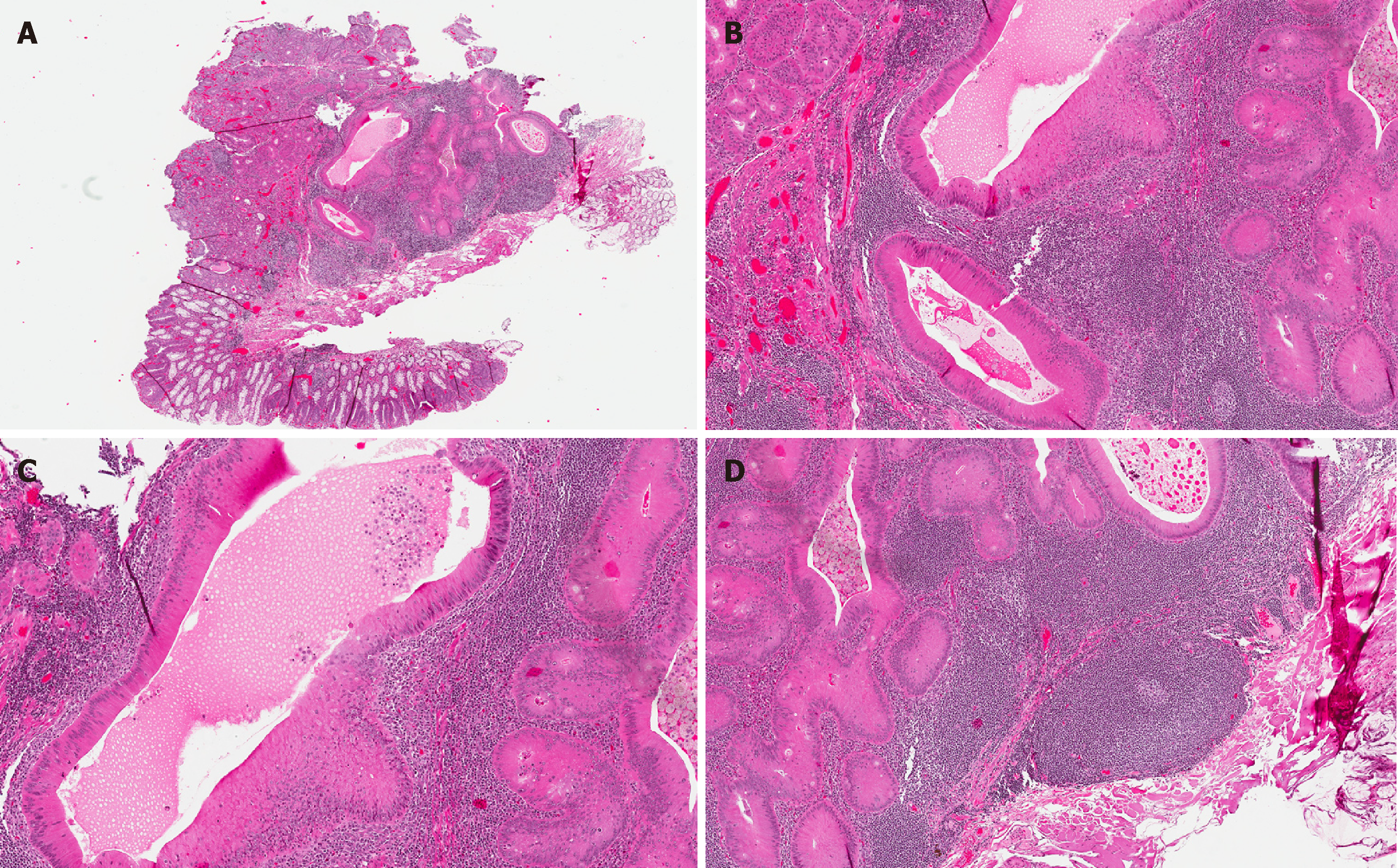
Figure 1 Hematoxylin and eosin stains.
A: Low power view of a gut- or gastrointestinal-associated lymphoid tissue/dome-type carcinoma showing a well-circumscribed submucosal nodular lesion composed of cystic glands seemingly embedded in a rich lymphoid matrix; B: The cells lining the cystic glands are columnar with eosinophilic, oncocytic cytoplasm displaying minimal cytological atypia and lacking goblet cells; C: The lumina of the cystic glands are distended by non-necrotic eosinophilic material; D: The very prominent lymphoid component is organized into follicles with reactive germinal centers.
- Citation: McCarthy AJ, Chetty R. Gut-associated lymphoid tissue or so-called “dome” carcinoma of the colon: Review. World J Gastrointest Oncol 2019; 11(1): 59-70
- URL: https://www.wjgnet.com/1948-5204/full/v11/i1/59.htm
- DOI: https://dx.doi.org/10.4251/wjgo.v11.i1.59


