Copyright
©The Author(s) 2017.
World J Gastrointest Endosc. Jun 16, 2017; 9(6): 282-295
Published online Jun 16, 2017. doi: 10.4253/wjge.v9.i6.282
Published online Jun 16, 2017. doi: 10.4253/wjge.v9.i6.282
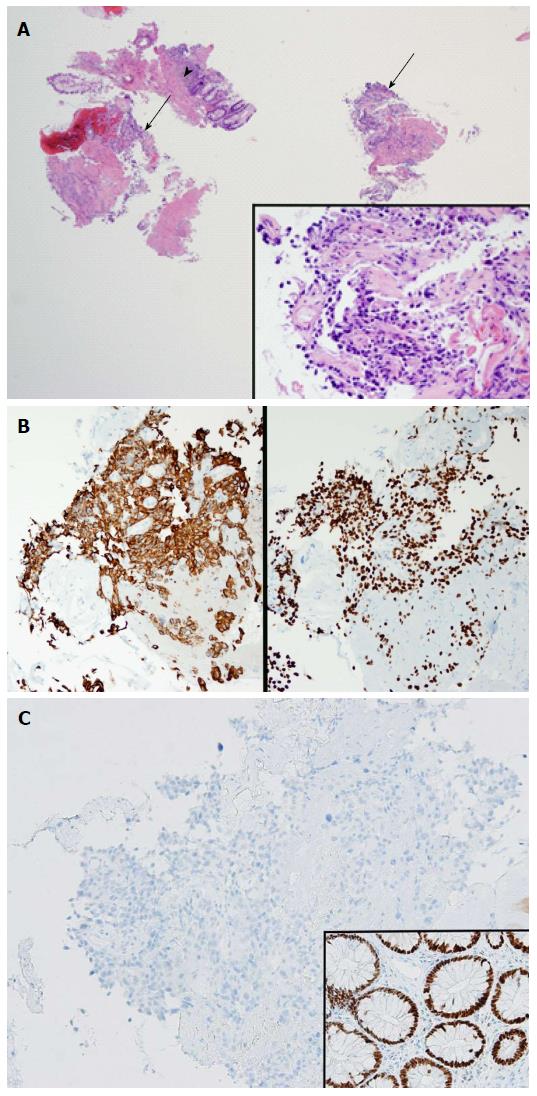
Figure 3 Histologic examination and immunohistochemical analysis.
A: Low power photomicrograph shows segments of detached poorly differentiated cancer (arrows), amidst segments of normal rectal mucosa (arrowhead). Right inset shows high power photomicrograph of polygonal tumor cells (HE stain) that have a histologic appearance characteristic for urothelial carcinoma. Immunohistochemistry confirmed the urothelial histology of the cancer (Figure 3B and C); B: Left: Immunohistochemistry for cytokeratin-20 demonstrates positive staining of tumor cytoplasm. Right: Immunohistochemistry for GATA-3 demonstrates positive staining of tumor cell nuclei. This immunohistochemical profile indicates that the rectal mass is of urothelial origin; C: Immunohistochemistry for CDX-2 shows negative staining of tumor cell nuclei, indicating that this tumor is not colonic adenocarcinoma. Right inset is a control of normal colonic glands set on the same slide which demonstrates positive nuclear staining for CDX-2.
- Citation: Aneese AM, Manuballa V, Amin M, Cappell MS. Bladder urothelial carcinoma extending to rectal mucosa and presenting with rectal bleeding. World J Gastrointest Endosc 2017; 9(6): 282-295
- URL: https://www.wjgnet.com/1948-5190/full/v9/i6/282.htm
- DOI: https://dx.doi.org/10.4253/wjge.v9.i6.282


