Copyright
©The Author(s) 2017.
World J Gastrointest Endosc. Jun 16, 2017; 9(6): 282-295
Published online Jun 16, 2017. doi: 10.4253/wjge.v9.i6.282
Published online Jun 16, 2017. doi: 10.4253/wjge.v9.i6.282
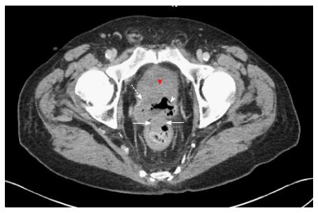
Figure 1 Abdominopelvic computed tomography angiography demonstrating thickened bladder wall (red arrowhead), with adjacent prostate margin (dashed arrow).
Air-filled cavity within the prostate gland (white arrowhead), is consistent with the known bladder urothelial carcinoma penetrating, via the prostate, into the rectum (horizontal solid arrows). The colonoscopic findings (Figure 2) strongly support this mechanism of cancer spread.
- Citation: Aneese AM, Manuballa V, Amin M, Cappell MS. Bladder urothelial carcinoma extending to rectal mucosa and presenting with rectal bleeding. World J Gastrointest Endosc 2017; 9(6): 282-295
- URL: https://www.wjgnet.com/1948-5190/full/v9/i6/282.htm
- DOI: https://dx.doi.org/10.4253/wjge.v9.i6.282


