Copyright
©The Author(s) 2016.
World J Gastrointest Endosc. Jan 25, 2016; 8(2): 67-76
Published online Jan 25, 2016. doi: 10.4253/wjge.v8.i2.67
Published online Jan 25, 2016. doi: 10.4253/wjge.v8.i2.67
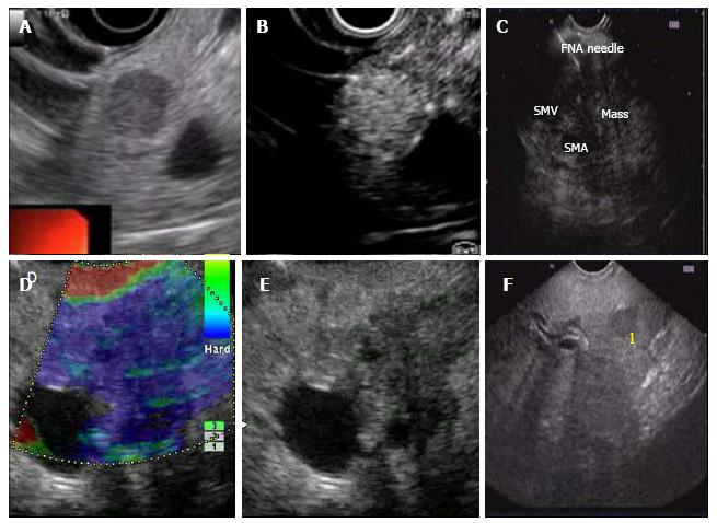
Figure 2 Contrast enhanced endoscopic ultrasound and endoscopic ultrasound elastography.
A and B: Neuroendocrine tumor in the head of pancreas (HOP) before (A) and after (B) contrast administration; C: Fine needle aspiration (FNA) of mass in the HOP; D and E: Carcinoma HOP, EUS elastographic (D) appearance and B mode EUS appearance (E); F: Carcinoma HOP with metastasis (1) in the left lobe of liver.
- Citation: Puri R, Manrai M, Thandassery RB, Alfadda AA. Endoscopic ultrasound in the diagnosis and management of carcinoma pancreas. World J Gastrointest Endosc 2016; 8(2): 67-76
- URL: https://www.wjgnet.com/1948-5190/full/v8/i2/67.htm
- DOI: https://dx.doi.org/10.4253/wjge.v8.i2.67


