Copyright
©The Author(s) 2016.
World J Gastrointest Endosc. Nov 16, 2016; 8(19): 690-696
Published online Nov 16, 2016. doi: 10.4253/wjge.v8.i19.690
Published online Nov 16, 2016. doi: 10.4253/wjge.v8.i19.690
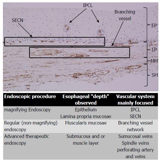
Figure 4 The figure shows the histology of a non-pathologic esophageal specimen.
The vessels’ wall has been colored by CD34, showing superficially the IPCLs (upper part of the lamina propria, arising the epithelium) and the SECN; deeply in the lamina propria the branching vessels. In the sumucosal layer also the drainage veins are evident. The table summarizes the vascular system observed and its own esophageal layer according to the different endoscopic procedure performed. SECN: Sub-epithelial capillary network; IPCL: Intrapapillary capillary loop; EP: Epithelium; LP: Lamina propria; MM: Muscolaris mucosa; SM: Submucosa.
- Citation: Maselli R, Inoue H, Ikeda H, Onimaru M, Yoshida A, Santi EG, Sato H, Hayee B, Kudo SE. Microvasculature of the esophagus and gastroesophageal junction: Lesson learned from submucosal endoscopy. World J Gastrointest Endosc 2016; 8(19): 690-696
- URL: https://www.wjgnet.com/1948-5190/full/v8/i19/690.htm
- DOI: https://dx.doi.org/10.4253/wjge.v8.i19.690


