Copyright
©The Author(s) 2015.
World J Gastrointest Endosc. May 16, 2015; 7(5): 563-566
Published online May 16, 2015. doi: 10.4253/wjge.v7.i5.563
Published online May 16, 2015. doi: 10.4253/wjge.v7.i5.563
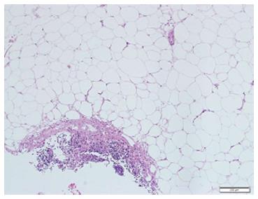
Figure 2 Mesenteric biopsy.
Mesenteric biopsy showing fibrotic band of dense collagen infiltrated by mixed inflammatory cells (lymphocytes, plasma cells and neutrophils). There is fat necrosis, no vasculitis or malignancy seen. There is no cellular atypia or lipoblast identified in the biopsy.
- Citation: Alhazzani W, Al-Shamsi HO, Greenwald E, Radhi J, Tse F. Chronic abdominal pain secondary to mesenteric panniculitis treated successfully with endoscopic ultrasonography-guided celiac plexus block: A case report. World J Gastrointest Endosc 2015; 7(5): 563-566
- URL: https://www.wjgnet.com/1948-5190/full/v7/i5/563.htm
- DOI: https://dx.doi.org/10.4253/wjge.v7.i5.563


