Copyright
©The Author(s) 2015.
World J Gastrointest Endosc. Mar 16, 2015; 7(3): 192-205
Published online Mar 16, 2015. doi: 10.4253/wjge.v7.i3.192
Published online Mar 16, 2015. doi: 10.4253/wjge.v7.i3.192
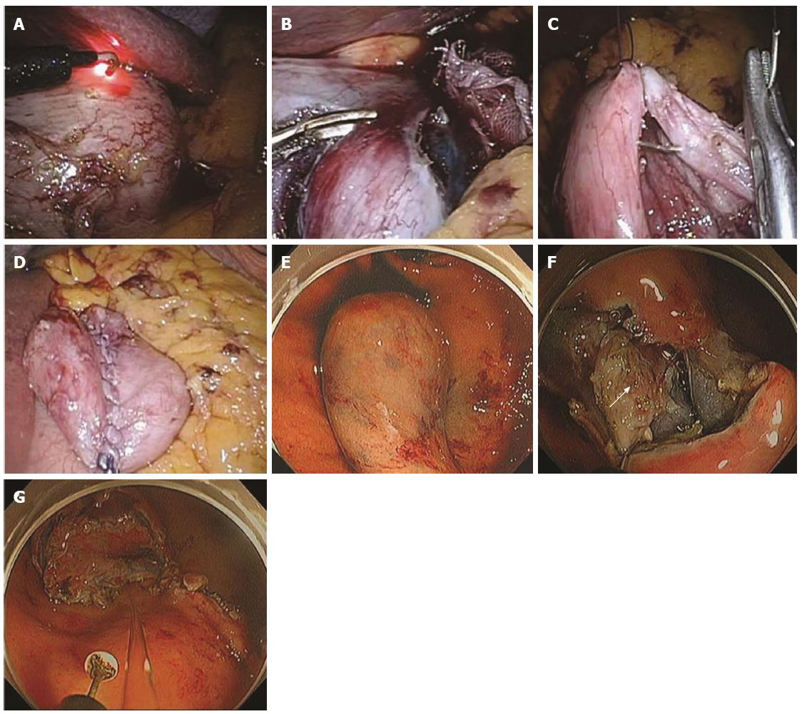
Figure 8 Procedure for non-exposed wall-inversion surgery.
A: Laparoscopic markings on the serosal surface guided by light from the fiber-optic probe shining through the gastric endoscope; B: Circumferential seromuscular layer dissection outside the serosal markings; C: Seromuscular layer suture closure; D: Laparoscopic view of inversion of the dissected area; E: Endoscopic view of massive protrusion of the inverted tissue; F: Serosal surface (arrow) identified during mucosubmucosal layer dissection; G: Flipped tissue to be resected (adopted from Mitsui et al[61]).
- Citation: Kim HH. Endoscopic treatment for gastrointestinal stromal tumor: Advantages and hurdles. World J Gastrointest Endosc 2015; 7(3): 192-205
- URL: https://www.wjgnet.com/1948-5190/full/v7/i3/192.htm
- DOI: https://dx.doi.org/10.4253/wjge.v7.i3.192


