Copyright
©The Author(s) 2015.
World J Gastrointest Endosc. Mar 16, 2015; 7(3): 192-205
Published online Mar 16, 2015. doi: 10.4253/wjge.v7.i3.192
Published online Mar 16, 2015. doi: 10.4253/wjge.v7.i3.192
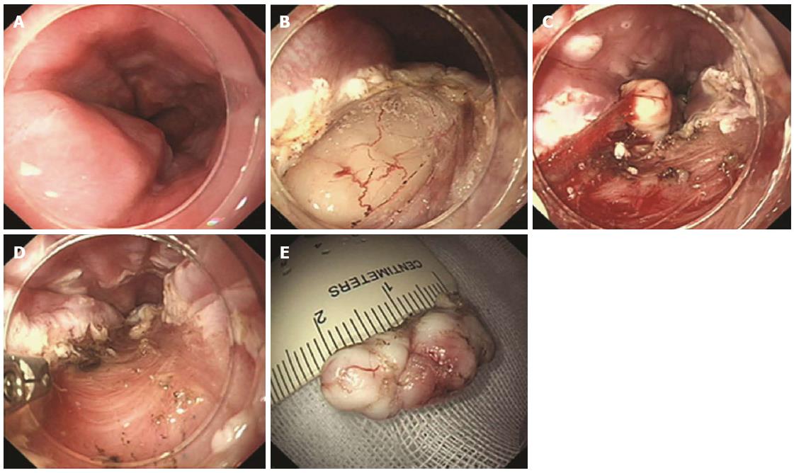
Figure 4 Endoscopic muscularis dissection of an esophageal subepithelial tumor originating from the proper muscle layers.
A: Endoscopic view of the esophageal submucosal tumor; B: Exposure of the tumor using a longitudinal incision; C: Blunt dissection of the tumor as deep as the proper muscle layer with a transparent hood; D: Stopping bleeding after blunt dissection; E: The whole tumor was removed F: Linear clipping was performed to close the submucosal entry site (adopted from Liu et al[44]).
- Citation: Kim HH. Endoscopic treatment for gastrointestinal stromal tumor: Advantages and hurdles. World J Gastrointest Endosc 2015; 7(3): 192-205
- URL: https://www.wjgnet.com/1948-5190/full/v7/i3/192.htm
- DOI: https://dx.doi.org/10.4253/wjge.v7.i3.192


