Copyright
©2013 Baishideng Publishing Group Co.
World J Gastrointest Endosc. May 16, 2013; 5(5): 265-269
Published online May 16, 2013. doi: 10.4253/wjge.v5.i5.265
Published online May 16, 2013. doi: 10.4253/wjge.v5.i5.265
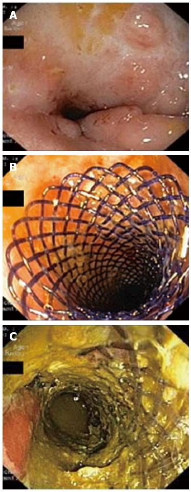
Figure 2 Endoscopic images.
A: Stricture of the distal sigmoid colon before stent insertion. The surrounding mucosa show no signs of active inflammation and few inflammatory polyps; B: Biodegradable stent deployed in the stricture at the end of the procedure; C: Endoscopic view 1 mo after the stent placement. The stent fibers present a translucent appearance and partial fragmentation.
- Citation: Rodrigues C, Oliveira A, Santos L, Pires E, Deus J. Biodegradable stent for the treatment of a colonic stricture in Crohn’s disease. World J Gastrointest Endosc 2013; 5(5): 265-269
- URL: https://www.wjgnet.com/1948-5190/full/v5/i5/265.htm
- DOI: https://dx.doi.org/10.4253/wjge.v5.i5.265


