Copyright
©2013 Baishideng Publishing Group Co.
World J Gastrointest Endosc. Nov 16, 2013; 5(11): 540-550
Published online Nov 16, 2013. doi: 10.4253/wjge.v5.i11.540
Published online Nov 16, 2013. doi: 10.4253/wjge.v5.i11.540
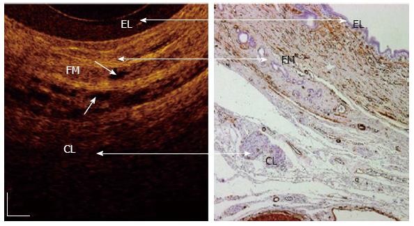
Figure 4 In vivo optical coherence tomography image of a normal sphincter of Oddi wall.
Three recognizable layers were observed from the surface of the duct to a depth of 1 mm (Color online). The inner single layers of epithelial (EL) cells are visible as a superficial, hypo-reflective layer (400-800 μm thick). The intermediate connective-muscular (FM) layer surrounding the epithelium is visible as a hyper-reflective layer (250-400 μm thick). The outer connective layer (CL) is visible as a hypo-reflective layer with longitudinal relatively hyper-reflective strips (smooth muscle fibers). Within intermediate and outer layer, vessels could be visualized (marked with arrows) as nonreflecting areas. The boundaries between the intermediate and outer layers are not clearly recognizable due to irregular distribution of the connective and muscular structure[59]. White scale bar: 150 μm.
- Citation: Mahmud MS, May GR, Kamal MM, Khwaja AS, Sun C, Vitkin A, Yang VX. Imaging pancreatobiliary ductal system with optical coherence tomography: A review. World J Gastrointest Endosc 2013; 5(11): 540-550
- URL: https://www.wjgnet.com/1948-5190/full/v5/i11/540.htm
- DOI: https://dx.doi.org/10.4253/wjge.v5.i11.540


