Copyright
©2012 Baishideng.
World J Gastrointest Endosc. Sep 16, 2012; 4(9): 387-397
Published online Sep 16, 2012. doi: 10.4253/wjge.v4.i9.387
Published online Sep 16, 2012. doi: 10.4253/wjge.v4.i9.387
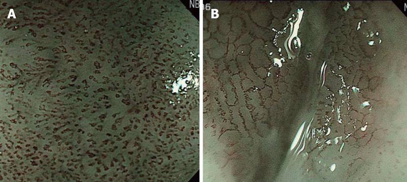
Figure 4 The development of narrow band imaging facilitated the visual observation of intra-epithelial papillary capillary loop, revolutionizing the diagnosis of the depth.
A: Findings of narrow band imaging-combined magnifying observation of a lesion diagnosed as M2 after endoscopic submucosal dissection (ESD). Dilated and curving intra-epithelial papillary capillary loop (IPCL) with an irregular width extending in a transverse direction; B: Findings of narrow band imaging-combined magnifying observation of a lesion diagnosed as M3 after ESD. IPCL was severely damaged , and a connection with the adjacent IPCL was also observed.
- Citation: Nonaka K, Nishimura M, Kita H. Role of narrow band imaging in endoscopic submucosal dissection. World J Gastrointest Endosc 2012; 4(9): 387-397
- URL: https://www.wjgnet.com/1948-5190/full/v4/i9/387.htm
- DOI: https://dx.doi.org/10.4253/wjge.v4.i9.387


