Copyright
©2012 Baishideng Publishing Group Co.
World J Gastrointest Endosc. Jul 16, 2012; 4(7): 335-338
Published online Jul 16, 2012. doi: 10.4253/wjge.v4.i7.335
Published online Jul 16, 2012. doi: 10.4253/wjge.v4.i7.335
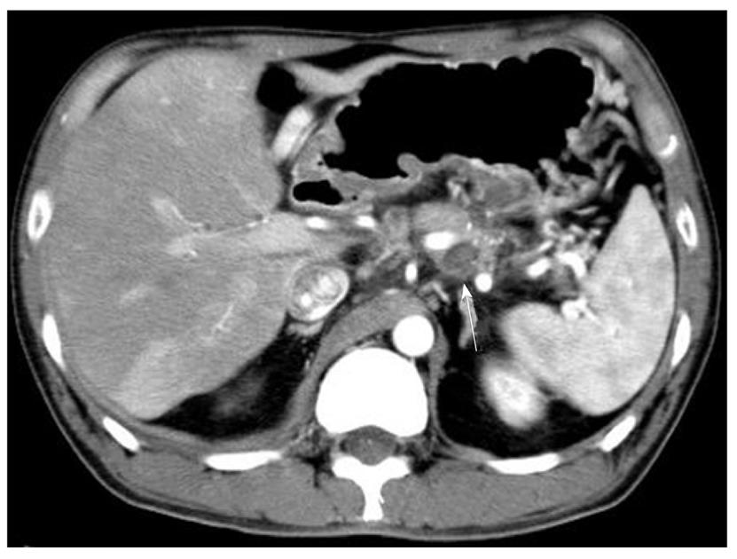
Figure 1 Abdominal computed tomographic findings.
A severely atrophied pancreas with a calcified body was noted. The pseudocyst (arrow) ranged from the back of the pancreas to the left mediastinum and was adjacent to the splenic artery.
- Citation: Fukatsu K, Ueda K, Maeda H, Yamashita Y, Itonaga M, Mori Y, Moribata K, Shingaki N, Deguchi H, Enomoto S, Inoue I, Maekita T, Iguchi M, Tamai H, Kato J, Ichinose M. A case of chronic pancreatitis in which endoscopic ultrasonography was effective in the diagnosis of a pseudoaneurysm. World J Gastrointest Endosc 2012; 4(7): 335-338
- URL: https://www.wjgnet.com/1948-5190/full/v4/i7/335.htm
- DOI: https://dx.doi.org/10.4253/wjge.v4.i7.335


