Copyright
©2011 Baishideng Publishing Group Co.
World J Gastrointest Endosc. Nov 16, 2011; 3(11): 231-234
Published online Nov 16, 2011. doi: 10.4253/wjge.v3.i11.231
Published online Nov 16, 2011. doi: 10.4253/wjge.v3.i11.231
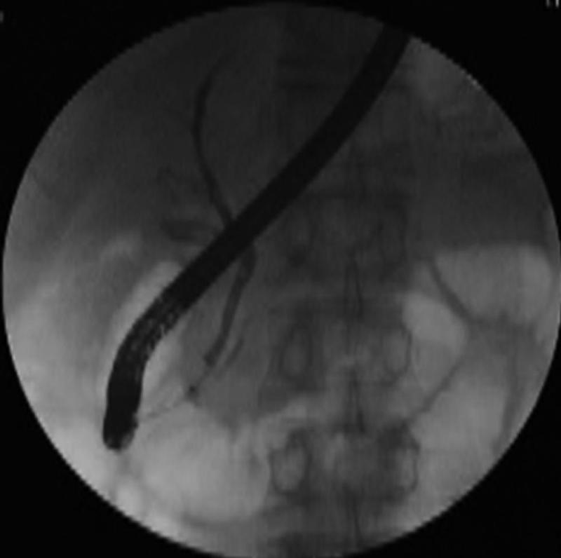
Figure 1 Pancreatic calculus is observed in the pancreatic body.
Although the pancreatic duct was imaged to some extent by endoscopic retrograde cholangiopancreatography, the catheter was dislodged due to the strong mobility of the duodenal papilla, after which only the bile duct was imaged.
- Citation: Sakai Y, Ishihara T, Tsuyuguchi T, Tawada K, Saito M, Kurosawa J, Tamura R, Togo S, Mikata R, Tada M, Yokosuka O. New cannulation method for pancreatic duct cannulation-bile duct guidewire-indwelling method. World J Gastrointest Endosc 2011; 3(11): 231-234
- URL: https://www.wjgnet.com/1948-5190/full/v3/i11/231.htm
- DOI: https://dx.doi.org/10.4253/wjge.v3.i11.231


