Copyright
©2010 Baishideng.
World J Gastrointest Endosc. Jan 16, 2010; 2(1): 15-19
Published online Jan 16, 2010. doi: 10.4253/wjge.v2.i1.15
Published online Jan 16, 2010. doi: 10.4253/wjge.v2.i1.15
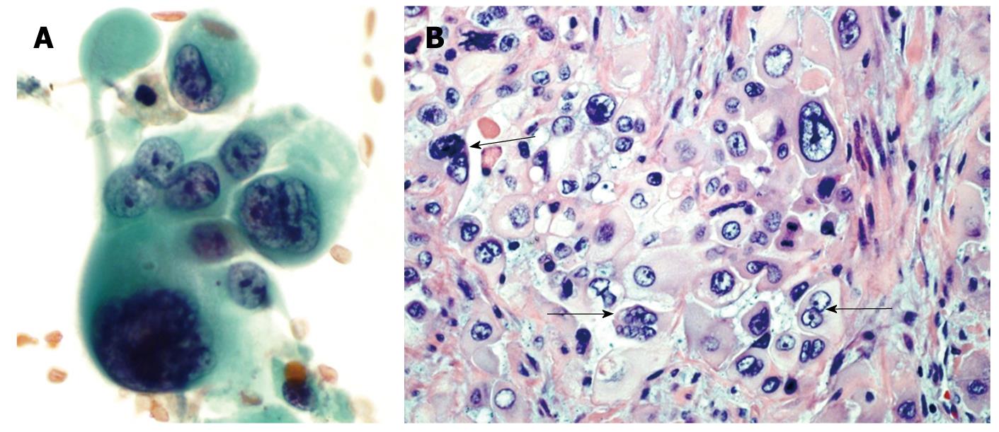
Figure 2 Pleomorphic giant cell tumor of pancreas.
A: Endoscopic-ultrasound guided fine needle aspirate of a pancreatic mass. These tumors are characterized by a mixture of markedly atypical mono and multinucleated giant cells. Both the mononuclear and multinucleated giant cells have similar appearing anaplastic nuclei (Papanicolaou stain); B: A tissue section from the pancreatectomy specimen. The neoplasm contains multinucleated giant cells (arrows) with hyperchromatic large, bizarre irregular tumor cell nuclei similar to the atypical mononuclear cells in the background (HE stain).
- Citation: Moore JC, Bentz JS, Hilden K, Adler DG. Osteoclastic and pleomorphic giant cell tumors of the pancreas: A review of clinical, endoscopic, and pathologic features. World J Gastrointest Endosc 2010; 2(1): 15-19
- URL: https://www.wjgnet.com/1948-5190/full/v2/i1/15.htm
- DOI: https://dx.doi.org/10.4253/wjge.v2.i1.15


