Copyright
©2010 Baishideng.
World J Gastrointest Endosc. Jan 16, 2010; 2(1): 15-19
Published online Jan 16, 2010. doi: 10.4253/wjge.v2.i1.15
Published online Jan 16, 2010. doi: 10.4253/wjge.v2.i1.15
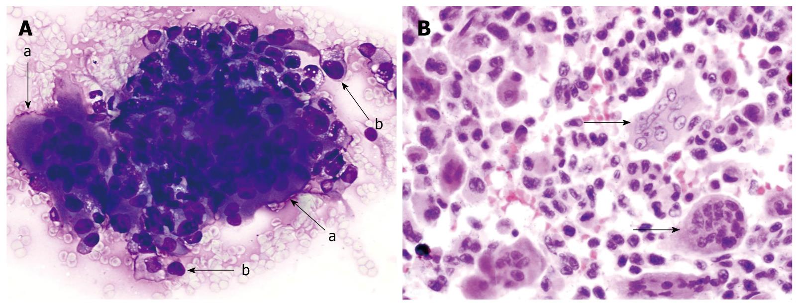
Figure 1 Osteoclastic giant cell tumor of pancreas.
A: Endoscopic-ultrasound guided fine needle aspirate specimen of a pancreatic mass. The smear slide contains a loose admixture of osteoclastic-giant cells admixed with oval to polygonal mononuclear cells (a-arrow). The individual giant cells have multiple bland appearing nuclei. The pleomorphic mononuclear cells are usually more numerous than the giant cells (b-arrow). (Romanowsky stain); B: A tissue section from the pancreatectomy specimen. The neoplasm contains a scattering of osteoclastic multinucleated giant cells with bland nuclei (arrows). These giant cells lie in a background of atypical mononuclear cells with pleomorphic nuclei. (HE stain).
- Citation: Moore JC, Bentz JS, Hilden K, Adler DG. Osteoclastic and pleomorphic giant cell tumors of the pancreas: A review of clinical, endoscopic, and pathologic features. World J Gastrointest Endosc 2010; 2(1): 15-19
- URL: https://www.wjgnet.com/1948-5190/full/v2/i1/15.htm
- DOI: https://dx.doi.org/10.4253/wjge.v2.i1.15


