
















©The Author(s) 2025.
World J Gastrointest Endosc. Sep 16, 2025; 17(9): 110424
Published online Sep 16, 2025. doi: 10.4253/wjge.v17.i9.110424
Published online Sep 16, 2025. doi: 10.4253/wjge.v17.i9.110424
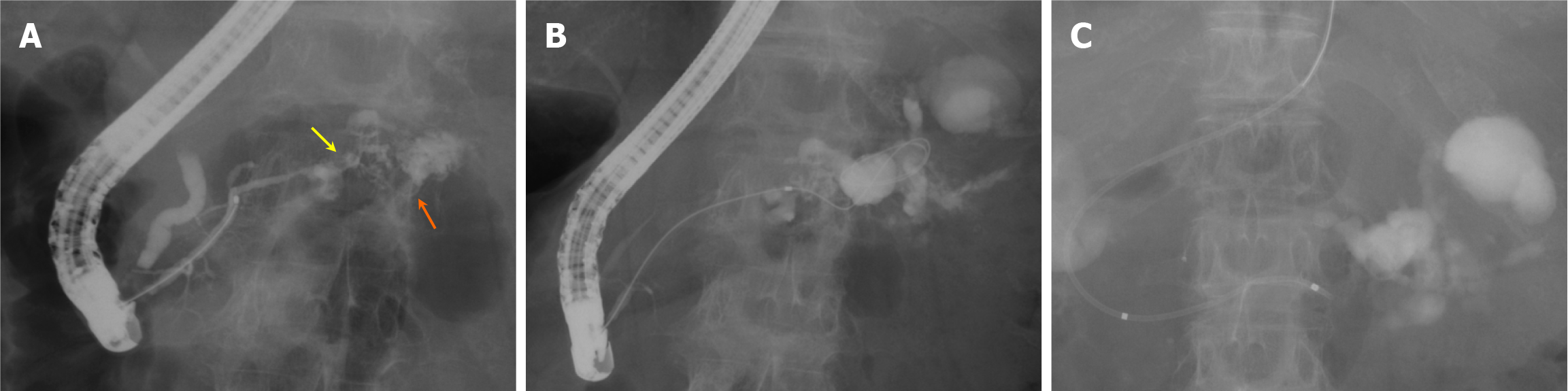
Figure 3 Findings from the initial endoscopic retrograde cholangiopancreatography.
A: Endoscopic retrograde cholangiopancreatography (ERCP) reveals an irregular stenosis of the main pancreatic duct in the pancreatic body. Distal to the stenosis, the main ductal structure is nearly obliterated by the tumor, and contrast medium is observed leaking into the area of pancreatic fluid leakage through several fine, disrupted ductal structures; B: Guidewire insertion into the leakage site was challenging even with multiple guidewires, but eventually, the guidewire was successfully advanced through an extremely fine tract (indicated by the yellow arrow in Figure 3A), which was not the main contrast-filled route (indicated by the orange arrow in Figure 3A). Attempts to dilate this trans-tumoral passage using a bougie dilator and a balloon dilator were unsuccessful; C: A 5-French endoscopic nasopancreatic drainage tube (Hanaco Medical Co., Saitama, Japan) is placed in the main pancreatic duct proximal to the stenosis.
- Citation: Makazu M, Koizumi K, Kubota J, Kimura K, Masuda S. Transpapillary drainage of pancreatic fluid leakage via a rigid trans-tumoral tract using a drill dilator: A case report. World J Gastrointest Endosc 2025; 17(9): 110424
- URL: https://www.wjgnet.com/1948-5190/full/v17/i9/110424.htm
- DOI: https://dx.doi.org/10.4253/wjge.v17.i9.110424