Copyright
©The Author(s) 2025.
World J Gastrointest Endosc. Jul 16, 2025; 17(7): 105773
Published online Jul 16, 2025. doi: 10.4253/wjge.v17.i7.105773
Published online Jul 16, 2025. doi: 10.4253/wjge.v17.i7.105773
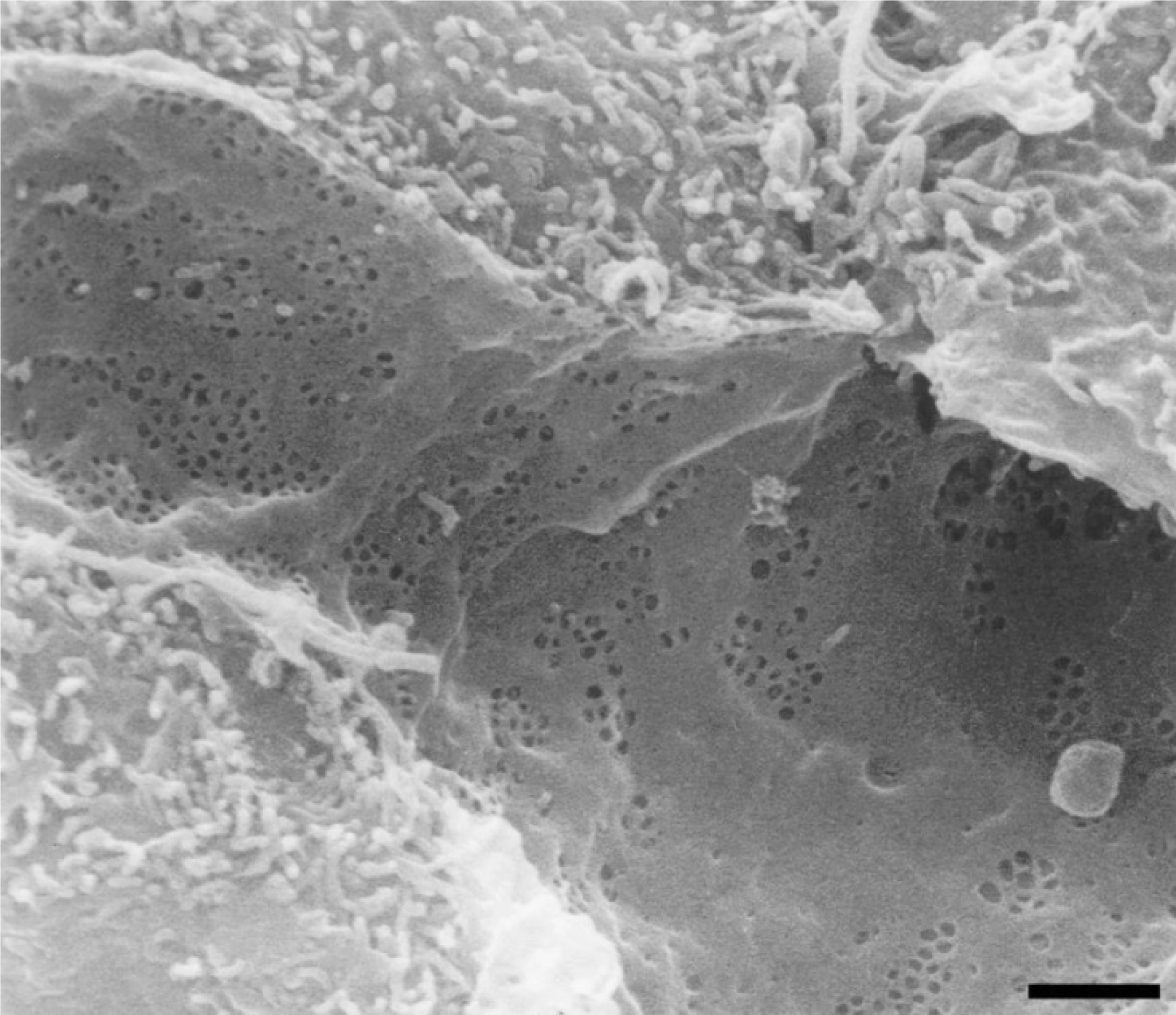
Figure 4 Low magnification scanning electron micrograph of the sinusoidal endothelium from a rat liver showing the fenestrated wall.
Shown is the clustering of fenestrae in sieve plates. Scale bar: 1 μm. Citation: Braet F, Wisse E. Structural and functional aspects of liver sinusoidal endothelial cell fenestrae: a review. Comp Hepatol 2002; 1(1): 1. Copyright ©The Author(s) 2019. Published by BioMed Central Ltd[7].
- Citation: Shin SY, Yeon HJ, Lee SO, Lee JR, Leem G, Han SJ. Fatal air embolism during intestinal endoscopy in Kasai portoenterostomy for biliary atresia: A case report. World J Gastrointest Endosc 2025; 17(7): 105773
- URL: https://www.wjgnet.com/1948-5190/full/v17/i7/105773.htm
- DOI: https://dx.doi.org/10.4253/wjge.v17.i7.105773


