Copyright
©The Author(s) 2022.
World J Gastrointest Endosc. Apr 16, 2022; 14(4): 191-204
Published online Apr 16, 2022. doi: 10.4253/wjge.v14.i4.191
Published online Apr 16, 2022. doi: 10.4253/wjge.v14.i4.191
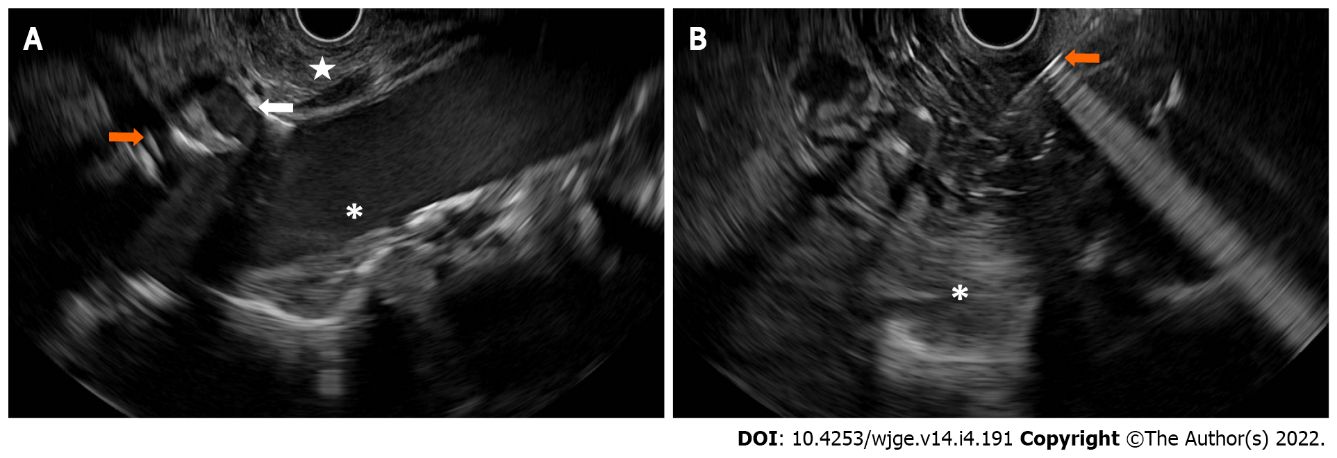
Figure 3 Endoscopic ultrasound guided celiac plexus neurolysis.
A: The structures while the echoendoscope is located at the posterior proximal gastric body/gastric cardia. A star demonstrates the pre-celiac region. The white arrow demonstrates the celiac trunk. A orange arrow demonstrates the superior mesenteric artery. An asterisk indicates the descending abdominal aorta; B: An area of hyperchoic blush of injected dehydrated alcohol (asterisk) delivered from a 19-gauge needle (arrow) for celiac plexus neurolysis.
- Citation: Kerdsirichairat T, Shin EJ. Endoscopic ultrasound guided interventions in the management of pancreatic cancer. World J Gastrointest Endosc 2022; 14(4): 191-204
- URL: https://www.wjgnet.com/1948-5190/full/v14/i4/191.htm
- DOI: https://dx.doi.org/10.4253/wjge.v14.i4.191


