Copyright
©The Author(s) 2019.
World J Gastrointest Endosc. Dec 16, 2019; 11(12): 548-560
Published online Dec 16, 2019. doi: 10.4253/wjge.v11.i12.548
Published online Dec 16, 2019. doi: 10.4253/wjge.v11.i12.548
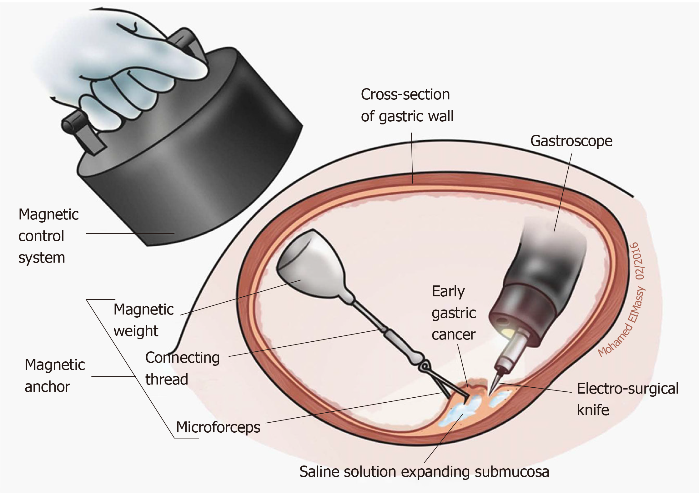
Figure 6 Application diagram of magnetic anchor guided endoscopic submucosal dissection[56].
A small internal permanent magnet is attached to the edge of a partially dissected lesion, while a large external permanent magnet is applied for retraction (an electromagnetic control system can also be selected). Used with permission from Baishideng Publishing Group Inc.
- Citation: Hu B, Ye LS. Endoscopic applications of magnets for the treatment of gastrointestinal diseases. World J Gastrointest Endosc 2019; 11(12): 548-560
- URL: https://www.wjgnet.com/1948-5190/full/v11/i12/548.htm
- DOI: https://dx.doi.org/10.4253/wjge.v11.i12.548


