Copyright
©The Author(s) 2018.
World J Gastrointest Endosc. Dec 16, 2018; 10(12): 378-382
Published online Dec 16, 2018. doi: 10.4253/wjge.v10.i12.378
Published online Dec 16, 2018. doi: 10.4253/wjge.v10.i12.378
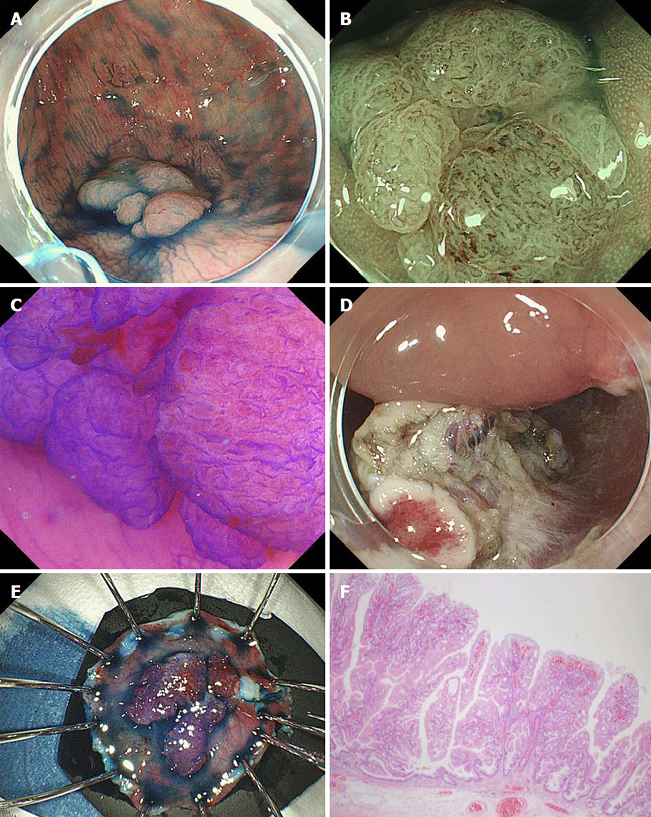
Figure 1 Endoscopic submucosal dissection of recurrent lesion in the cecum.
A: A Local recurrence (laterally spreading tumor, granular type) was identified in the cecum 18 mo after piecemeal endoscopic mucosal resection; B: The Japan Narrow-band imaging Expert Team classification was type 2B[19]; C: Kudo’s pit pattern was VI[20]. The laterally spreading tumor was diagnosed as an intramucosal lesion and ESD performed; D, E: Although there was severe fibrosis in the submucosal layer, en bloc resection was achieved; F: The pathological diagnosis was adenocarcinoma arising from a sessile serrated adenoma/polyp, type 0-IIa, 16 × 15 mm, pTis, pHM0, pVM0; ER0, Cur EA; pap > tub1, ly0, v0.
- Citation: Shichijo S, Takeuchi Y, Uedo N, Ishihara R. Management of local recurrence after endoscopic resection of neoplastic colonic polyps. World J Gastrointest Endosc 2018; 10(12): 378-382
- URL: https://www.wjgnet.com/1948-5190/full/v10/i12/378.htm
- DOI: https://dx.doi.org/10.4253/wjge.v10.i12.378


