Copyright
©The Author(s) 2017.
World J Gastrointest Endosc. Aug 16, 2017; 9(8): 346-358
Published online Aug 16, 2017. doi: 10.4253/wjge.v9.i8.346
Published online Aug 16, 2017. doi: 10.4253/wjge.v9.i8.346
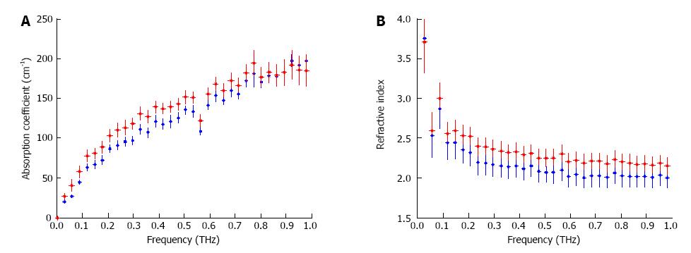
Figure 2 Terahertz spectroscopic results for the absorption coefficient (A) and refractive index (B) of fresh excisions of normal (blue) and cancerous (red) colon tissues[38] (Printed with permission).
THz: Terahertz.
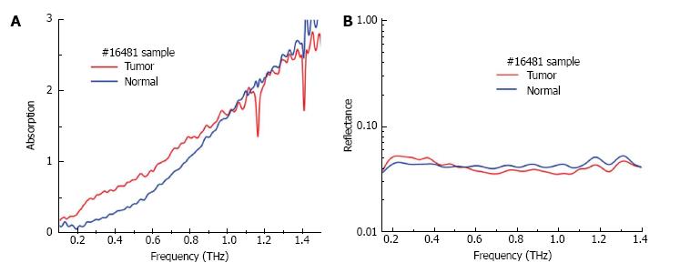
Figure 3 Absorption (A) and reflectance (B) measurements of paraffin embedded dehydrated fixed specimens of normal and cancerous colon tissue[39] (printed with permission).
THz: Terahertz.
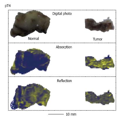
Figure 4 Photographs, absorption (transmittance) and reflection images of formalin fixed dehydrated colon tissue[39] (printed with permission).
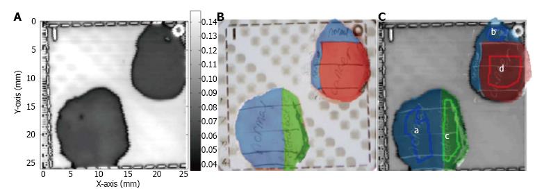
Figure 5 An example terahertz image of excised cancerous, dysplastic and healthy colonic tissues.
A: Example terahertz (THz) image of tissue containing healthy regions, dysplasia and cancerous tissue; B: The histology results (drawn onto a photographic image of the tissue samples); C: The histology results are overlaid on the THz image. In this example, regions a and b are normal tissue, c is dysplastic tissue and d is cancerous tissue[38] (printed with permission).
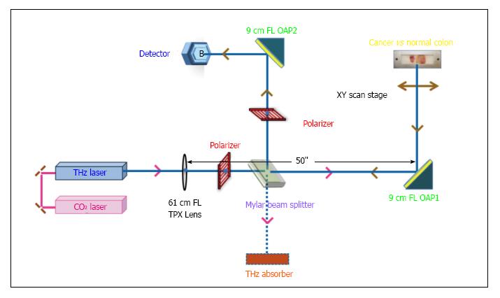
Figure 6 Schematic of continuous-wave terahertz reflection imaging system[42].
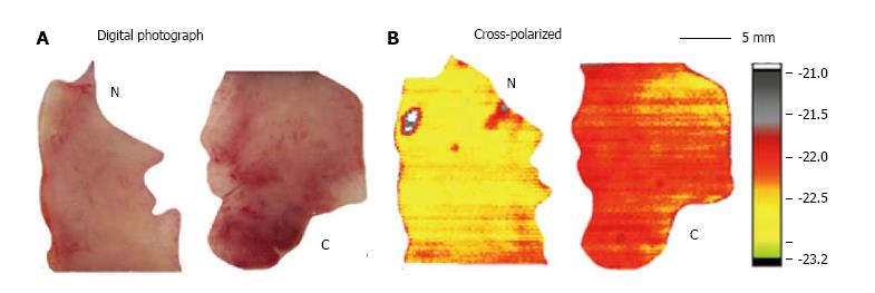
Figure 7 Digital photograph (A) and corresponding terahertz reflectance images (B) of normal (N) and cancerous (C) colon tissue[42].
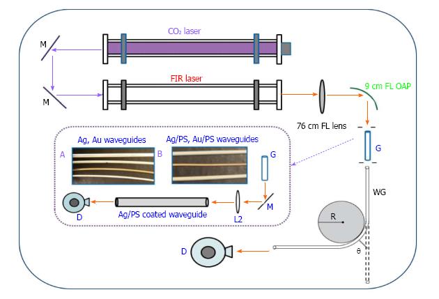
Figure 8 Experimental setup for the transmission loss measurement in metal and metal dielectric.
Inset: A: 4 mm Ag (top), 3 mm Ag, 2 mm Au, and 2 mm Ag; B: 3 mm Ag/PS (top), 2 mm Au/PS, and 4 mm Ag/PS) coated terahertz waveguides[54].
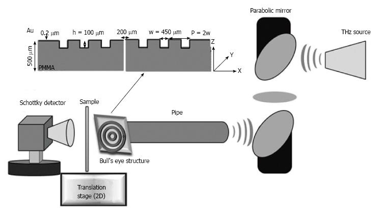
Figure 9 Schematic of waveguide based terahertz near-field transmission imaging system[41] (printed with permission).
PMMA: Polymethyl methacrylate.
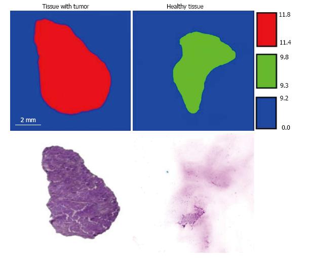
Figure 10 Terahertz transmittance images and stained histology sections showing cancerous and normal colon tissue[41] (printed with permission).
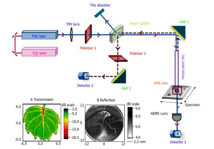
Figure 11 Schematic of single-channel prototype terahertz endoscopic imaging setup.
Inset: Terahertz (A) transmission imaging of a small 10 mm leaf, and (B) reflection imaging of a 25-cent coin. THz: Terahertz.
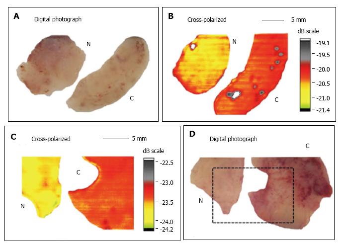
Figure 12 Digital photograph, cross-polarized terahertz reflection images of normal N vs cancerous C human colonic formalin fixed (A and B) and fresh (C and D) tissue sets[58].
- Citation: Doradla P, Joseph C, Giles RH. Terahertz endoscopic imaging for colorectal cancer detection: Current status and future perspectives. World J Gastrointest Endosc 2017; 9(8): 346-358
- URL: https://www.wjgnet.com/1948-5190/full/v9/i8/346.htm
- DOI: https://dx.doi.org/10.4253/wjge.v9.i8.346


