Copyright
©The Author(s) 2021.
World J Gastrointest Endosc. Sep 16, 2021; 13(9): 356-370
Published online Sep 16, 2021. doi: 10.4253/wjge.v13.i9.356
Published online Sep 16, 2021. doi: 10.4253/wjge.v13.i9.356
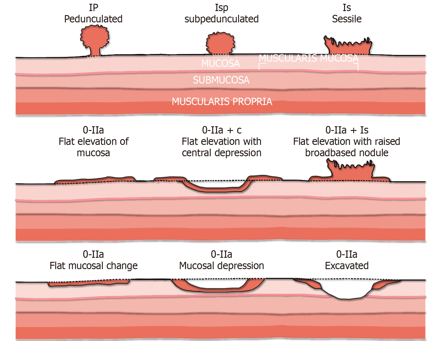
Figure 1 The Paris endoscopic classification of colorectal polyps.
Adapted from[23].
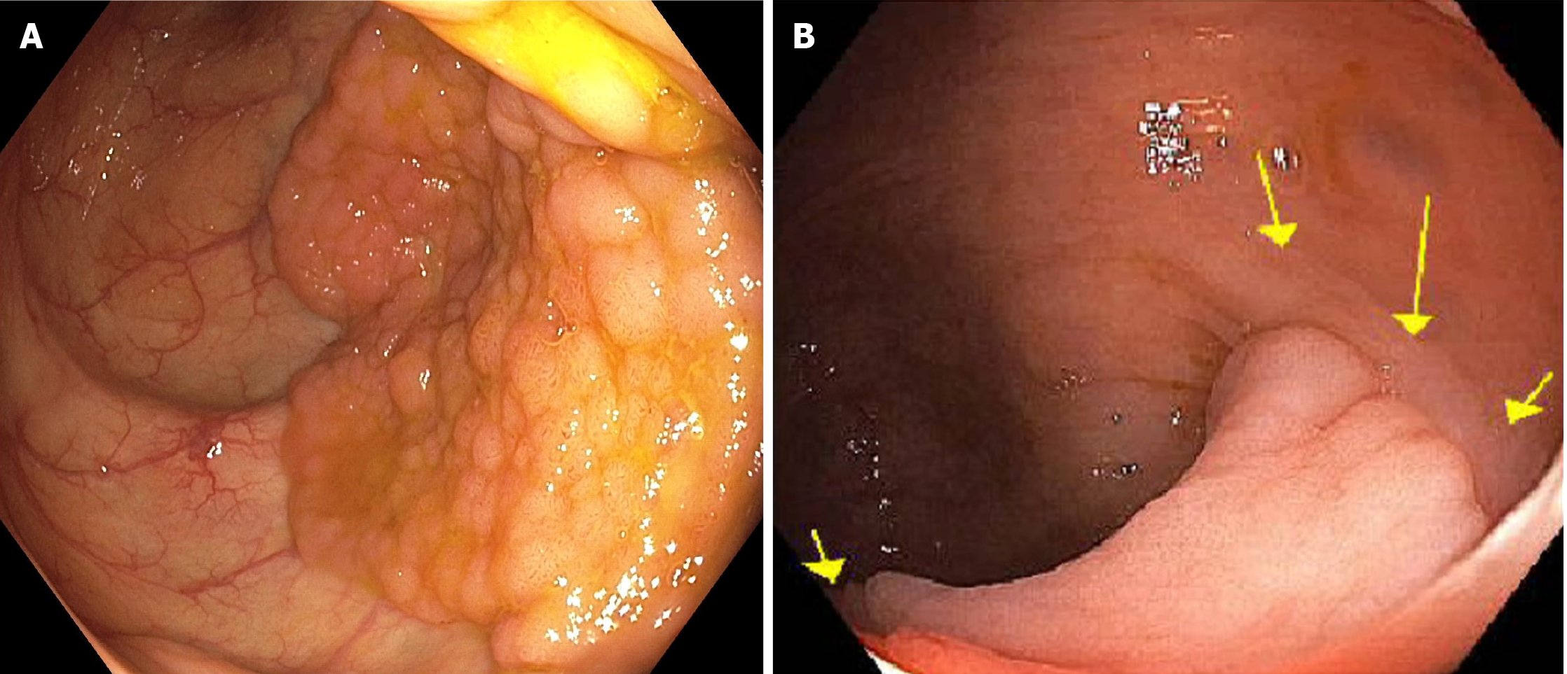
Figure 2 Lateral spreading tumor.
A: Lateral spreading tumor with granular surface; B: Lateral spreading tumor non-granular type highlighted by arrows.
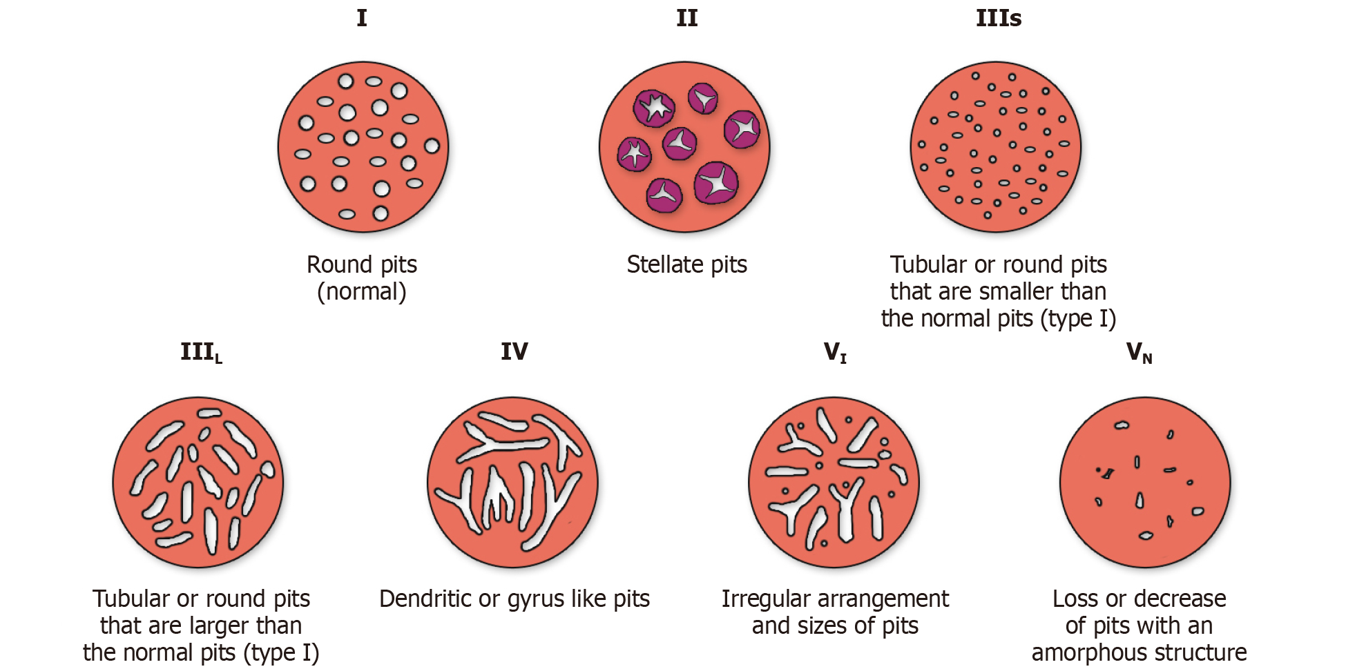
Figure 3 Kudo classification of pit pattern (Adapted from Kudo et al[33]).
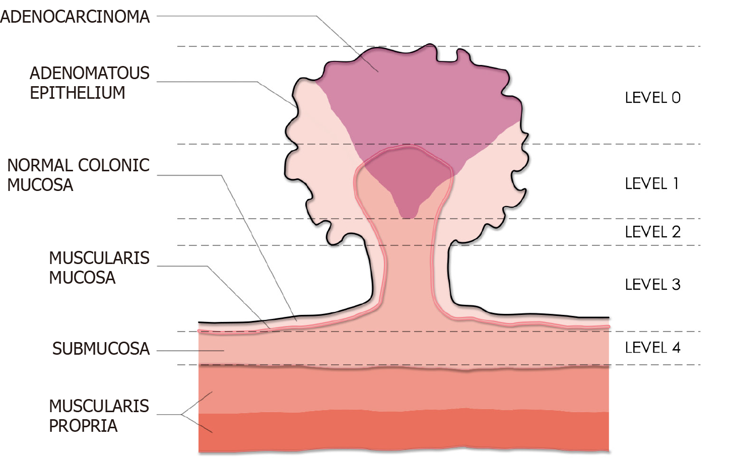
Figure 4 Haggitt classification system of pedunculated polyps (Adapted from Haggitt et al[35]).
This system categorizes polyps into five levels (level 0 to 4) based on the degree of invasion. In this illustration, an adenocarcinoma confined to the head of the polyp would be classified as Level 1.
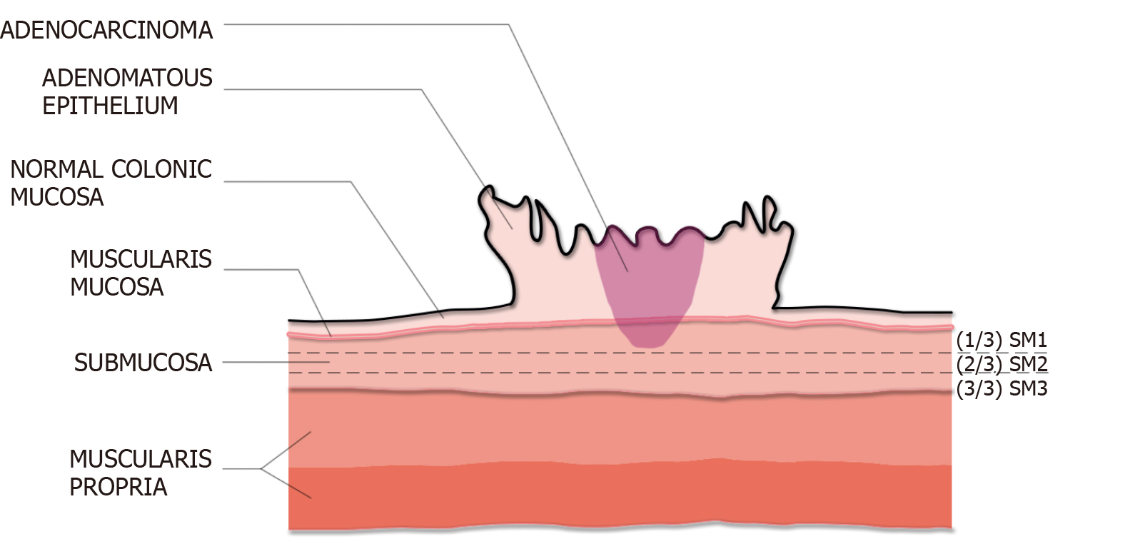
Figure 5 Kudo and Kikuchi classification (adapted from Kikuchi et al[37]).
Depth of submucosal invasion is divided into Sm1 (invasion into the upper third of the submucosa), Sm2 (invasion into the middle third), Sm3 (invasion into the lower third). In this illustration, the adenocarcinoma is a superficial lesion with Sm1 invasion.
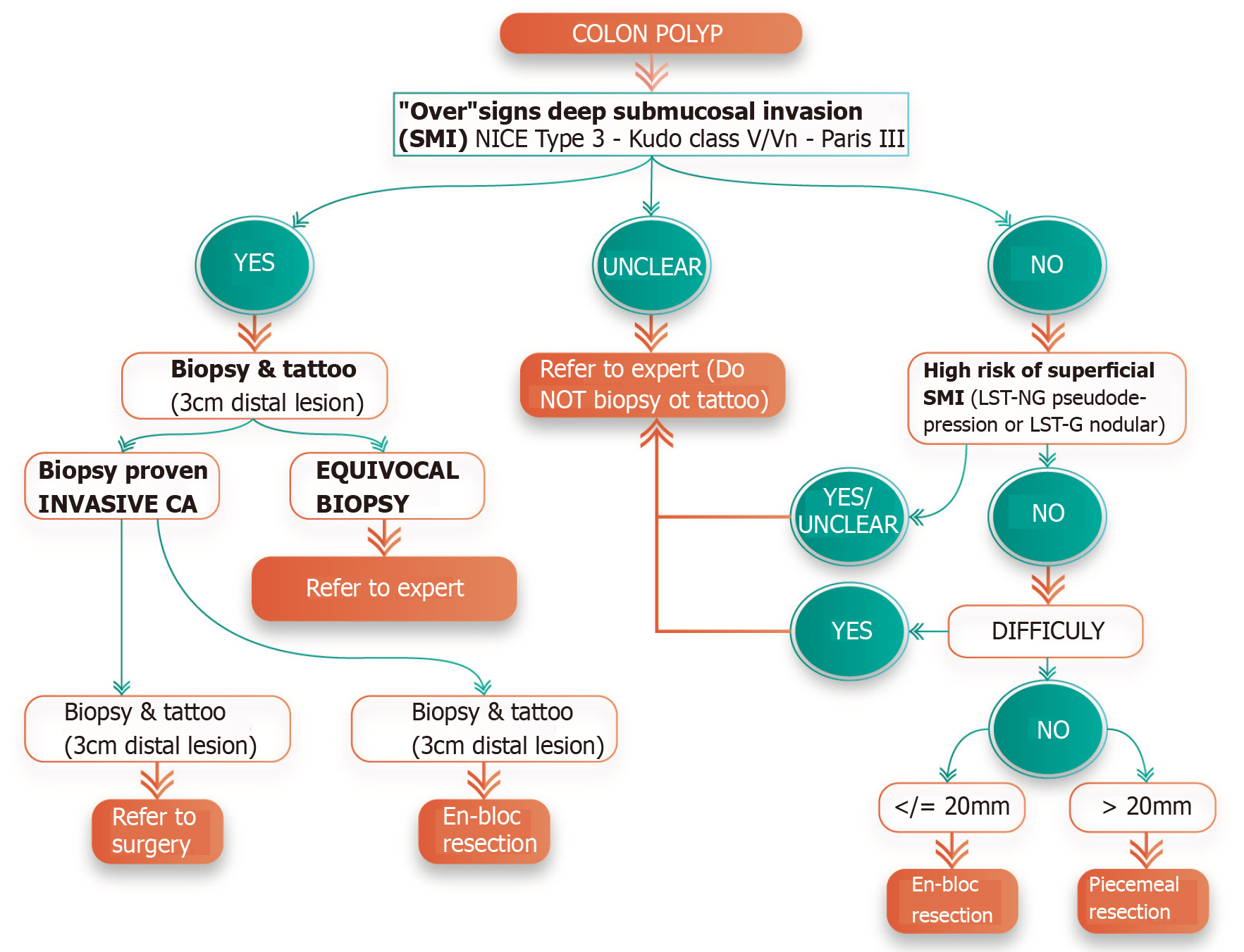
Figure 6 Decision tree algorithm for the evaluation and management of colorectal polyps.
- Citation: Mathews AA, Draganov PV, Yang D. Endoscopic management of colorectal polyps: From benign to malignant polyps. World J Gastrointest Endosc 2021; 13(9): 356-370
- URL: https://www.wjgnet.com/1948-5190/full/v13/i9/356.htm
- DOI: https://dx.doi.org/10.4253/wjge.v13.i9.356


