
















©The Author(s) 2017.
World J Hepatol. Dec 28, 2017; 9(36): 1372-1377
Published online Dec 28, 2017. doi: 10.4254/wjh.v9.i36.1372
Published online Dec 28, 2017. doi: 10.4254/wjh.v9.i36.1372
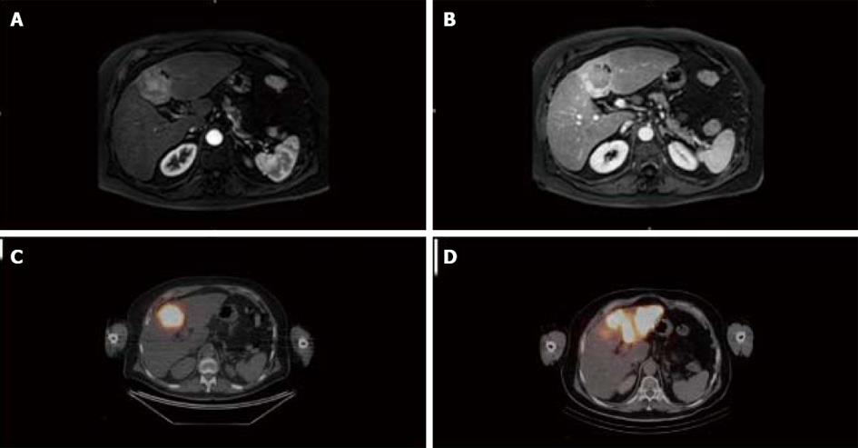
Figure 1 Preoperative imaging.
A and B: Baseline contrast-enhanced magnetic resonance imaging (MRI). Contrast-enhanced MRI demonstrated a 40 mm mass in segment IV of the liver with arterial wash-in (A) and wash-out on the portal venous phase (B) and features of cirrhosis (irregular surface, relative hypertrophy of segment I); C and D: Selective intra-tumor deposition of 90Y microspheres after first SIRT session (C) and deposition of 90Y microspheres to segments II and III after the second SIRT session (D).
- Citation: Vouche M, Degrez T, Bouazza F, Delatte P, Galdon MG, Hendlisz A, Flamen P, Donckier V. Sequential tumor-directed and lobar radioembolization before major hepatectomy for hepatocellular carcinoma. World J Hepatol 2017; 9(36): 1372-1377
- URL: https://www.wjgnet.com/1948-5182/full/v9/i36/1372.htm
- DOI: https://dx.doi.org/10.4254/wjh.v9.i36.1372