Copyright
©The Author(s) 2017.
World J Hepatol. Dec 28, 2017; 9(36): 1361-1366
Published online Dec 28, 2017. doi: 10.4254/wjh.v9.i36.1361
Published online Dec 28, 2017. doi: 10.4254/wjh.v9.i36.1361
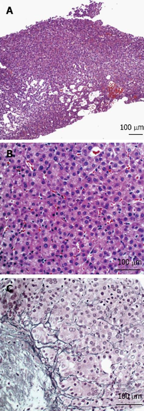
Figure 2 Ultrasound-guided liver biopsy.
A: Biopsy of the mass shows a solid growth pattern of hepatocytes (H+E, 100 ×); B: Neoplastic hepatocytes contain hyperchromatic, pleomorphic and enlarged nuclei (H+E, 200 ×); C: Reticulin stain demonstrates thickened trabeculae and decreased staining in the lesion (200 ×).
- Citation: Torres-Landa S, Muñoz-Abraham AS, Fortune BE, Gurung A, Pollak J, Emre SH, Rodriguez-Davalos MI, Schilsky ML. De-novo hepatocellular carcinoma after pediatric living donor liver transplantation. World J Hepatol 2017; 9(36): 1361-1366
- URL: https://www.wjgnet.com/1948-5182/full/v9/i36/1361.htm
- DOI: https://dx.doi.org/10.4254/wjh.v9.i36.1361


