Copyright
©The Author(s) 2017.
World J Hepatol. Jul 8, 2017; 9(19): 840-849
Published online Jul 8, 2017. doi: 10.4254/wjh.v9.i19.840
Published online Jul 8, 2017. doi: 10.4254/wjh.v9.i19.840
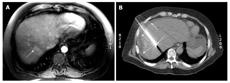
Figure 6 Computed tomography guided radiofrequency ablation in a 56-year-old lady with colorectal liver metastases.
A: Axial post gadolinium T1 weighted magnetic resonance image shows a 2.7 cm (arrow) hepatic dome metastases; B: The radiofrequency ablation was performed with the patient in supine position and needle placement through the anterolateral intercostal approach. Hydrodissection was performed in this patient (arrows).
- Citation: Kambadakone A, Baliyan V, Kordbacheh H, Uppot RN, Thabet A, Gervais DA, Arellano RS. Imaging guided percutaneous interventions in hepatic dome lesions: Tips and tricks. World J Hepatol 2017; 9(19): 840-849
- URL: https://www.wjgnet.com/1948-5182/full/v9/i19/840.htm
- DOI: https://dx.doi.org/10.4254/wjh.v9.i19.840


