Copyright
©The Author(s) 2017.
World J Hepatol. Jul 8, 2017; 9(19): 840-849
Published online Jul 8, 2017. doi: 10.4254/wjh.v9.i19.840
Published online Jul 8, 2017. doi: 10.4254/wjh.v9.i19.840
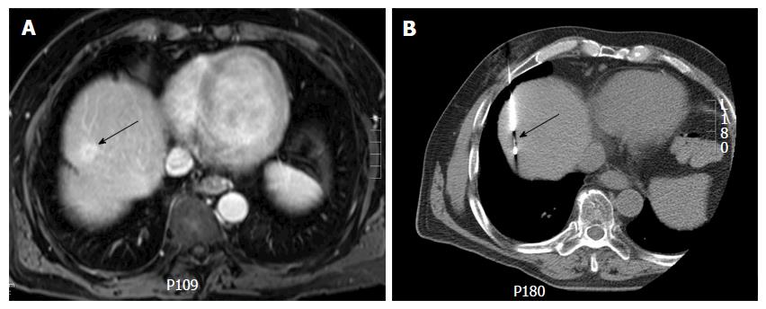
Figure 2 Computed tomography guided biopsy of a liver dome lesion in a 61-year-old man.
A: Axial post gadolinium T1-weighted magnetic resonance image shows a 2 cm lesion (arrow) in the hepatic dome. On pre-procedural computed tomography, the tumor was not well seen and contrast could not be administered due to iodine allergy; B: Needle placement for biopsy was done based on use of anatomic landmarks (arrow) (configuration of inferior vena cava, cardiac margin and aorta) via a transpulmonary approach. Histopathology: Hepatocellular carcinoma.
- Citation: Kambadakone A, Baliyan V, Kordbacheh H, Uppot RN, Thabet A, Gervais DA, Arellano RS. Imaging guided percutaneous interventions in hepatic dome lesions: Tips and tricks. World J Hepatol 2017; 9(19): 840-849
- URL: https://www.wjgnet.com/1948-5182/full/v9/i19/840.htm
- DOI: https://dx.doi.org/10.4254/wjh.v9.i19.840


