Copyright
©The Author(s) 2017.
World J Hepatol. Jun 8, 2017; 9(16): 752-756
Published online Jun 8, 2017. doi: 10.4254/wjh.v9.i16.752
Published online Jun 8, 2017. doi: 10.4254/wjh.v9.i16.752
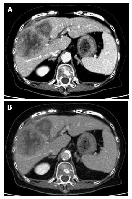
Figure 1 Contrast enhanced computed tomography scan showed a tumor located in the right anterior compartment of the liver with extrahepatic hematoma.
The tumors were hyperattenuating relative to the noncancerous liver parenchyma on arterial-phase (A) and hypo- or isoattenuating on delayed phase (B).
- Citation: Akabane S, Ban T, Kouriki S, Tanemura H, Nakazaki H, Nakano M, Shinozaki N. Successful surgical resection of ruptured cholangiolocellular carcinoma: A rare case of a primary hepatic tumor. World J Hepatol 2017; 9(16): 752-756
- URL: https://www.wjgnet.com/1948-5182/full/v9/i16/752.htm
- DOI: https://dx.doi.org/10.4254/wjh.v9.i16.752


