Copyright
©The Author(s) 2017.
World J Hepatol. May 18, 2017; 9(14): 645-656
Published online May 18, 2017. doi: 10.4254/wjh.v9.i14.645
Published online May 18, 2017. doi: 10.4254/wjh.v9.i14.645
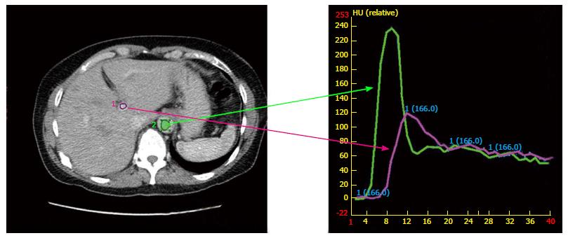
Figure 1 Image of a time-density intravenous contrast enhancement called Dynamic Contrast Enhanced computerized tomography.
Images are binned by location in respiratory cycle and when the contrast density within a vessel (such as the aorta and portal vein) signifies the non-contrast, arterial and wash-out phase. Time is measured in seconds and density is measured in Hounsefield units (HU).
- Citation: Lock MI, Klein J, Chung HT, Herman JM, Kim EY, Small W, Mayr NA, Lo SS. Strategies to tackle the challenges of external beam radiotherapy for liver tumors. World J Hepatol 2017; 9(14): 645-656
- URL: https://www.wjgnet.com/1948-5182/full/v9/i14/645.htm
- DOI: https://dx.doi.org/10.4254/wjh.v9.i14.645


