Copyright
©The Author(s) 2016.
World J Hepatol. Jan 28, 2016; 8(3): 148-161
Published online Jan 28, 2016. doi: 10.4254/wjh.v8.i3.148
Published online Jan 28, 2016. doi: 10.4254/wjh.v8.i3.148
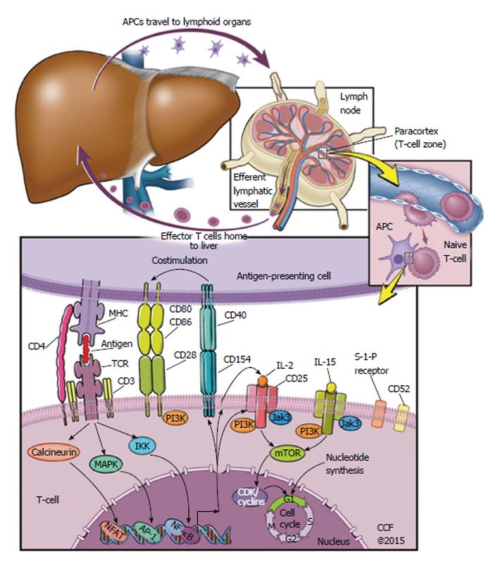
Figure 1 Signaling pathways targeted by modern immunosuppressive agents.
Starting from the top left, an antigen-presenting cell (APC) migrates from local tissue to lymphoid organs. In the paracortex of the lymphoid organ, the APC meets a naive T-cell. If the naive T-cell has a T-cell receptor (TCR) that binds the antigen as it is presented by a Major Histocompatibility Complex on the APC, a set of other T-cell-APC interactions are likely to follow. This includes T-cell CD4 binding to the MHC on an APC, as well as costimulatory signals via extracellular receptors CD28 or CD40. After this set of T-cell-APC interactions begins, a set of intracellular signals follow towards the nucleus of the T-cell. The naive T-cell is then activated, and begins to replicate. This replication is called clonal expansion, and produces a population of T-cells that eventually migrate back to the tissue that contains antigen. AP-1: Activator protein 1; CD: Cluster of differentiation; CDK: Cyclin-dependent kinase; IKK: Inhibitor of kappa-B kinase; IL: Interleukin; Jak3: Janus-associated kinase 3; PI3K: Phosphatidyl inositide 3-kinase; MAPK: Mitogen-activated protein kinase; mTOR: Mechanistic target of rapamycin; NFAT: Nuclear factor of activated T-cells; NF-κB: Nuclear factor kappa-light-chain-enhancer of activated B cells; TCR: T-cell receptor; MHC: Major histocompatibility complex.
- Citation: Ascha MS, Ascha ML, Hanouneh IA. Management of immunosuppressant agents following liver transplantation: Less is more. World J Hepatol 2016; 8(3): 148-161
- URL: https://www.wjgnet.com/1948-5182/full/v8/i3/148.htm
- DOI: https://dx.doi.org/10.4254/wjh.v8.i3.148


