Copyright
©The Author(s) 2015.
World J Hepatol. Apr 28, 2015; 7(6): 874-884
Published online Apr 28, 2015. doi: 10.4254/wjh.v7.i6.874
Published online Apr 28, 2015. doi: 10.4254/wjh.v7.i6.874
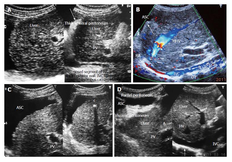
Figure 5 Ultrasonography showing ascites and evidence of chronic peritonitis in a patient with cirrhosis.
A: Acute exacerbation in a patient with liver cirrhosis showing hepatic portion of the inferior vena cava (IVC) filled with organizing thrombus and ascites with thick visceral peritoneum-indicating presence of bacterial peritonitis; B: Ultrasonography of a patient with liver cirrhosis due to HVCS: showing long segment stenosis of IVC with thick old organized thrombus (OT) along the posterior wall of the hepatic portion of the IVC. Note the presence of ascites (ASC) and irregular margin of the liver indicated by an arrow. Middle hepatic vein (MHV) is obstructed at its orifice and shows distal segmental stenosis; C: Ultrasonography of a patient with liver cirrhosis due to HVCS showing inferior vena cava stenosis, a calcified focus (c) in the liver and ascites; D: Ultrasonography of patient with liver cirrhosis due to HVCS showing IVC stenosis with organized thrombus on posterior wall and ASC and thick visceral peritoneum, suggestive of chronic bacterial peritonitis. PV: Portal vein; HVCS: Hepatic vena cava syndrome; RA: Right atrium; HV: Hepatic vein.
- Citation: Shrestha SM. Liver cirrhosis in hepatic vena cava syndrome (or membranous obstruction of inferior vena cava). World J Hepatol 2015; 7(6): 874-884
- URL: https://www.wjgnet.com/1948-5182/full/v7/i6/874.htm
- DOI: https://dx.doi.org/10.4254/wjh.v7.i6.874


