Copyright
©The Author(s) 2015.
World J Hepatol. Apr 18, 2015; 7(5): 814-818
Published online Apr 18, 2015. doi: 10.4254/wjh.v7.i5.814
Published online Apr 18, 2015. doi: 10.4254/wjh.v7.i5.814
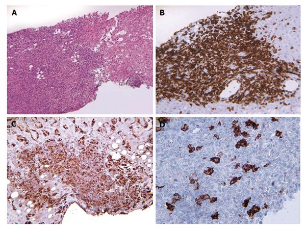
Figure 2 Second liver biopsy with diffuse mononuclear infiltrate (A, × 100) composed of predominantly small T lymphocytes (B, × 100) and histiocytes (C, × 200) with scattered large neoplastic B cells (CD20+) (D, × 400).
- Citation: Nosotti L, Baiocchini A, Bonifati C, Visco-Comandini U, Mirisola C, Del Nonno F. Unusual case of B cell lymphoma after immunosuppressive treatment for psoriasis. World J Hepatol 2015; 7(5): 814-818
- URL: https://www.wjgnet.com/1948-5182/full/v7/i5/814.htm
- DOI: https://dx.doi.org/10.4254/wjh.v7.i5.814


