Copyright
©The Author(s) 2015.
World J Hepatol. Mar 27, 2015; 7(3): 468-487
Published online Mar 27, 2015. doi: 10.4254/wjh.v7.i3.468
Published online Mar 27, 2015. doi: 10.4254/wjh.v7.i3.468
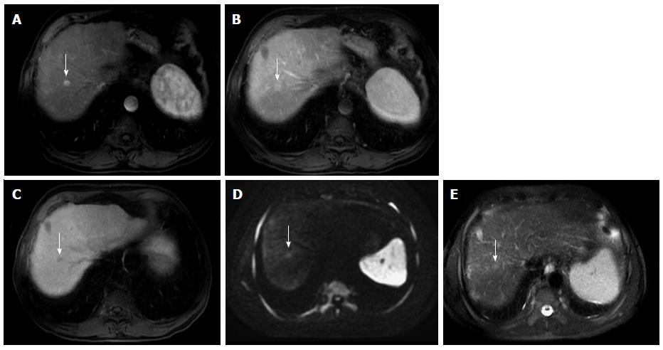
Figure 18 Small hepatocellular carcinoma showing the value of diffusion weighted image and hepatocyte-specific agents.
A: Post-contrast fat-suppressed 3D-GRE T1-weighted images during the (A) late hepatic arterial, (B) delayed, and (C) hepatobiliary phases; D: Diffusion weighted image (DWI) (b = 50); E: Fat-suppressed SSFSE T2-weighted image. There is a small right hepatic lobe nodule, which demonstrates increased arterial enhancement (arrow, A), fading out on the delayed images (arrow, B), and demonstrates significantly decreased uptake on the hepatobiliary phase (arrow, C). The lesion also demonstrates clear increased signal on DWI (arrow, D) and subtle increased T2 signal (arrow, E). The constellation of findings is consistent with HCC. The decreased uptake on the hepatobiliary phase and increased signal on DWI increased the accuracy of HCC diagnosis in this patient with a small hypervascular nodule. HCC: Hepatocellular carcinoma; GRE: Gradient recalled echo; SSFSE: Single-shot fast spin-echo.
- Citation: Watanabe A, Ramalho M, AlObaidy M, Kim HJ, Velloni FG, Semelka RC. Magnetic resonance imaging of the cirrhotic liver: An update. World J Hepatol 2015; 7(3): 468-487
- URL: https://www.wjgnet.com/1948-5182/full/v7/i3/468.htm
- DOI: https://dx.doi.org/10.4254/wjh.v7.i3.468


