Copyright
©The Author(s) 2015.
World J Hepatol. Mar 27, 2015; 7(3): 468-487
Published online Mar 27, 2015. doi: 10.4254/wjh.v7.i3.468
Published online Mar 27, 2015. doi: 10.4254/wjh.v7.i3.468
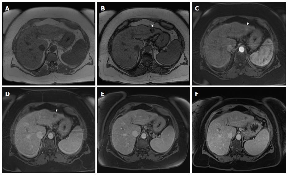
Figure 14 Slow growing, well-differentiated, fat-containing, small hepatocellular carcinoma.
In-phase (A) and opposed-phase GRE T1 weighted images (B); Post-contrast fat-suppressed 3D-GRE T1-weighted images during the late hepatic (C) arterial and delayed phases (D). E, F: Post-contrast fat-suppressed 3D-GRE T1-weighted delayed images from prior examinations performed 1 and 2 years prior, respectively. The is a small left hepatic lobe nodule, which demonstrates drop of signal on the opposed-phase image (arrowhead, B) compared to the in-phase image (A), increased arterial enhancement (arrowhead, C), and delayed washout (arrowhead,D). The lesion does not demonstrate significant change in size from the immediate prior examination (E). However, when compared with a more remote examination (F), substantial interval growth can be appreciated consistent with a slow growing, well-differentiated, fat-containing, small HCC. HCC: Hepatocellular carcinoma; GRE: Gradient recalled echo.
- Citation: Watanabe A, Ramalho M, AlObaidy M, Kim HJ, Velloni FG, Semelka RC. Magnetic resonance imaging of the cirrhotic liver: An update. World J Hepatol 2015; 7(3): 468-487
- URL: https://www.wjgnet.com/1948-5182/full/v7/i3/468.htm
- DOI: https://dx.doi.org/10.4254/wjh.v7.i3.468


