Copyright
©The Author(s) 2015.
World J Hepatol. Sep 8, 2015; 7(19): 2229-2236
Published online Sep 8, 2015. doi: 10.4254/wjh.v7.i19.2229
Published online Sep 8, 2015. doi: 10.4254/wjh.v7.i19.2229
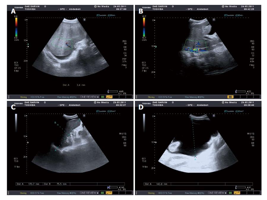
Figure 2 Ultrasound Doppler examination: Liver with signs of cirrhotic changes and luminal occlusion of hepatic veins to 3.
4 mm (A); Non-occlusive thrombosis of retrohepatic segment of the inferior vena cava (B); Signs of portal hypertension development: ascites (C) and splenomegaly (D).
- Citation: Reshetnyak TM, Seredavkina NV, Satybaldyeva MA, Nasonov EL, Reshetnyak VI. Liver transplantation in a patient with primary antiphospholipid syndrome and Budd-Chiari syndrome. World J Hepatol 2015; 7(19): 2229-2236
- URL: https://www.wjgnet.com/1948-5182/full/v7/i19/2229.htm
- DOI: https://dx.doi.org/10.4254/wjh.v7.i19.2229


