
















©The Author(s) 2015.
World J Hepatol. Aug 18, 2015; 7(17): 2069-2079
Published online Aug 18, 2015. doi: 10.4254/wjh.v7.i17.2069
Published online Aug 18, 2015. doi: 10.4254/wjh.v7.i17.2069
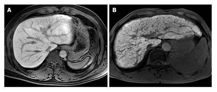
Figure 8 Fat saturated T1-weighted magnetic resonance images of 20-min delay after injection of gadoxetate disodium.
A: The image of healthy patient shows homogenous high signal intensity of hepatic parenchyma; B: The image of patient with liver cirrhosis shows heterogeneity of hepatic parenchyma enhancement with hypointense reticulations representing septal fibrosis, and decreased enhancement degree as compared with the image (A).
- Citation: Yeom SK, Lee CH, Cha SH, Park CM. Prediction of liver cirrhosis, using diagnostic imaging tools. World J Hepatol 2015; 7(17): 2069-2079
- URL: https://www.wjgnet.com/1948-5182/full/v7/i17/2069.htm
- DOI: https://dx.doi.org/10.4254/wjh.v7.i17.2069