Copyright
©2014 Baishideng Publishing Group Inc.
World J Hepatol. Aug 27, 2014; 6(8): 580-595
Published online Aug 27, 2014. doi: 10.4254/wjh.v6.i8.580
Published online Aug 27, 2014. doi: 10.4254/wjh.v6.i8.580
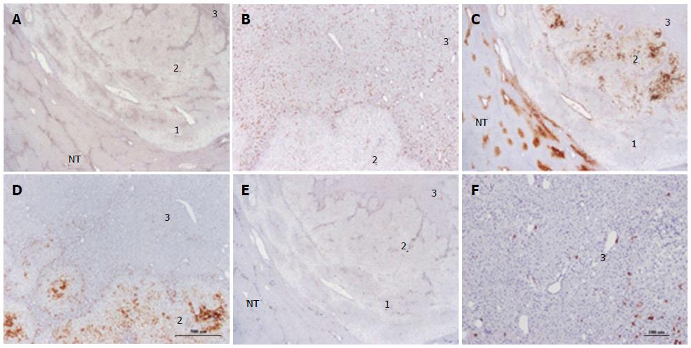
Figure 20 Unclassified hepatocellular adenoma.
Woman born in 1980; oral contraceptives for 6 years. BMI 22.2 kg/m2. Abdominal pain. Imaging: one nodule 3 cm, hepatocellular adenoma (HCA). Segmentectomy VI 2004. A, B: On CD34, three zones (1-3) are seen in this nodule. Zone 1 is the external limit of the nodule; zone 2 is intermediate and zone 3 represents the quasi totality of the nodule. Only zone 3 is diffusely positive. In zone 2, CD34 positivity is seen along vascular axis. C, D: Glutamine synthase (GS) staining: zone 3 is negative. Zone 1 is negative except around veins. In zone 2, GS staining is patchy. E, F: CK 7 - in zone 3, few cells, possibly progenitor cells are positive. In zone 2, positive cells are seen along vascular axis. This nodule has been classified as UHCA because all specific markers were negative. It is not rare to observe a thin peripheral rim which is CD34 negative /GS positive in unclassified HCA or β-HCA. In this case, the presence of 2 zones different from the bulk of the tumor remains unexplained but should not change the diagnosis.
- Citation: Sempoux C, Balabaud C, Bioulac-Sage P. Pictures of focal nodular hyperplasia and hepatocellular adenomas. World J Hepatol 2014; 6(8): 580-595
- URL: https://www.wjgnet.com/1948-5182/full/v6/i8/580.htm
- DOI: https://dx.doi.org/10.4254/wjh.v6.i8.580


