Copyright
©2014 Baishideng Publishing Group Inc.
World J Hepatol. Aug 27, 2014; 6(8): 580-595
Published online Aug 27, 2014. doi: 10.4254/wjh.v6.i8.580
Published online Aug 27, 2014. doi: 10.4254/wjh.v6.i8.580
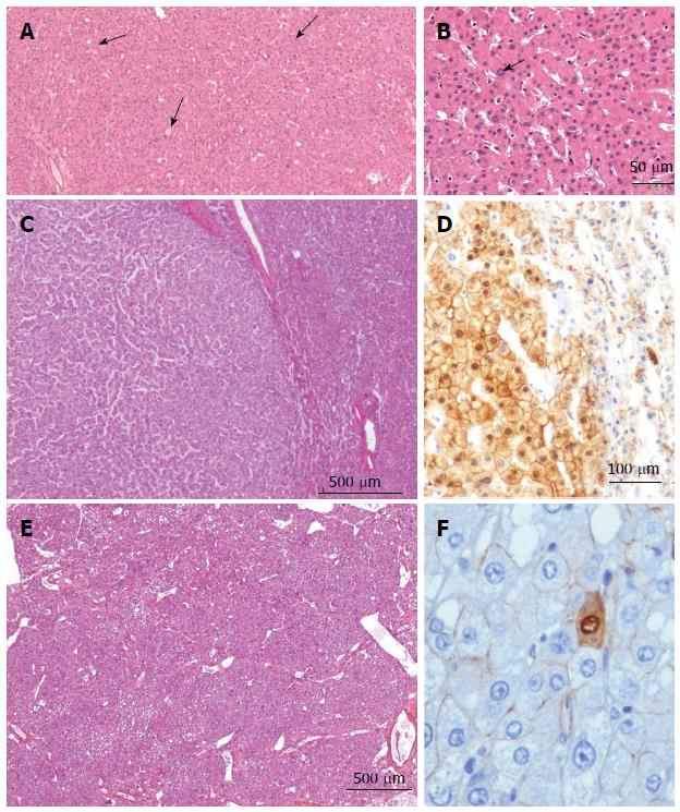
Figure 16 β-hepatocellular adenoma.
A, B: Man, 16 years old under androgen treatment for a hematological disorder. Imaging: several nodules; tumorectomy of one nodule 3 cm. HE: Hepatocellular adenoma (HCA) with some glandular arrangements ( arrowhead) and a few larger, irregular nuclei (arrow). According to the clinical context, the diagnosis of β-catenin is very likely. C, D: Woman born in 1953; oral contraceptives for 4 years. Danazol one year (endometriosis). BMI 19.5 kg/m2. Asthenia. Imaging: one nodule 3 cm. Right hepatectomy in 1989. C: HE: Well limited HCA with no features of H-HCA, or of IHCA. By default, the diagnosis of β-catenin is therefore a possibility. D: Aberrant cytoplasmic and nuclear expression of β-catenin in numerous hepatocytes confirms the diagnosis of β-HCA; E, F: Woman born in 1980; oral contraceptives for 12 years. BMI 20.4 kg/m2. Abdominal pain. Imaging: one nodule 15 cm: HCA. Right hepatectomy 2009. E: HE - numerous vessels within the HCA. This aspect seems to be quite characteristic of β-HCA and of some unclassified HCA; F: Aberrant expression of β-HCA in very few hepatocytes confirms the diagnosis of β-HCA.
- Citation: Sempoux C, Balabaud C, Bioulac-Sage P. Pictures of focal nodular hyperplasia and hepatocellular adenomas. World J Hepatol 2014; 6(8): 580-595
- URL: https://www.wjgnet.com/1948-5182/full/v6/i8/580.htm
- DOI: https://dx.doi.org/10.4254/wjh.v6.i8.580


