Copyright
©2014 Baishideng Publishing Group Inc.
World J Hepatol. Aug 27, 2014; 6(8): 580-595
Published online Aug 27, 2014. doi: 10.4254/wjh.v6.i8.580
Published online Aug 27, 2014. doi: 10.4254/wjh.v6.i8.580
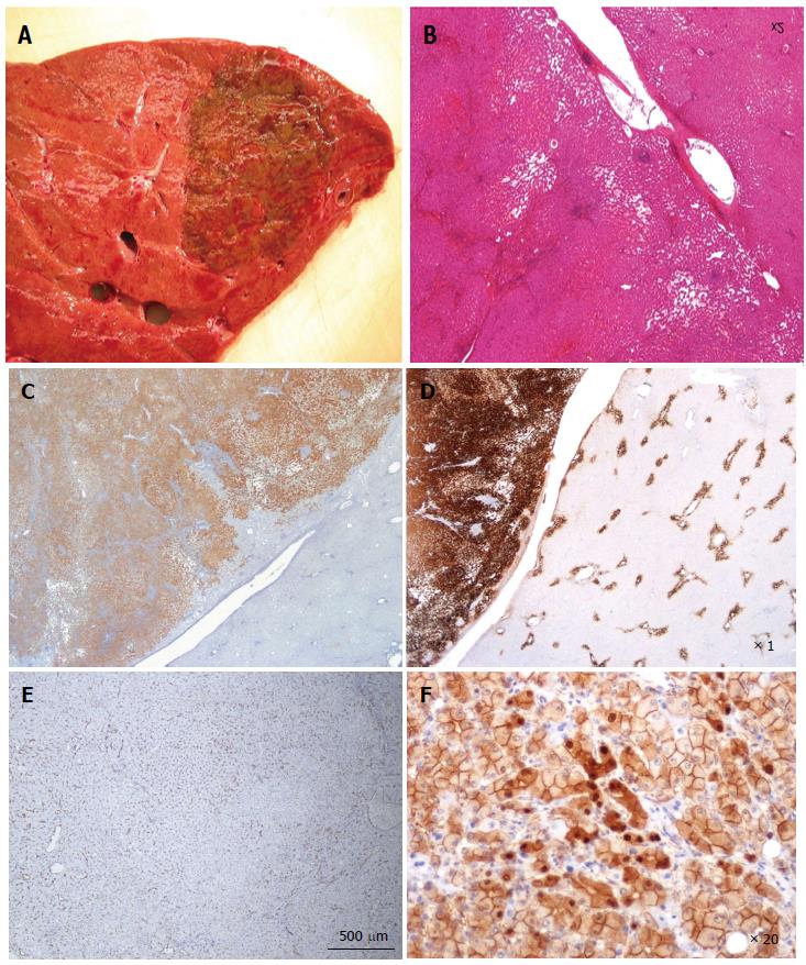
Figure 13 β-catenin activated, inflammatory hepatocellular adenoma.
A-F: Man born in 1971. BMI 21.6 kg/m2. By chance, discovery of one nodule 6 cm. Imaging: focal nodular hyperplasia. Right hepatectomy 2006. A: Fresh specimen: pigmented, irregular tumor. Non tumoral liver is normal. B: HE: Hepatocellular adenoma with sinusoidal dilatation and inflammatory infiltrate (on the left); large vessels at the junction with non tumoral liver. C: Diffuse expression of C-reactive protein by tumoral hepatocytes, with sharp demarcation from the non tumoral liver. D: Strong and diffuse glutamine synthase (GS) expression contrasting with normal staining of GS in adjacent non tumoral liver (in a few pericentrolobular hepatocytes). E: Large areas are positive for CD34, but not diffuse diffusely; F: Aberrant nuclear and cytoplasmic expression of β-catenin in quite numerous hepatocytes.
- Citation: Sempoux C, Balabaud C, Bioulac-Sage P. Pictures of focal nodular hyperplasia and hepatocellular adenomas. World J Hepatol 2014; 6(8): 580-595
- URL: https://www.wjgnet.com/1948-5182/full/v6/i8/580.htm
- DOI: https://dx.doi.org/10.4254/wjh.v6.i8.580


