Copyright
©2014 Baishideng Publishing Group Inc.
World J Hepatol. Aug 27, 2014; 6(8): 580-595
Published online Aug 27, 2014. doi: 10.4254/wjh.v6.i8.580
Published online Aug 27, 2014. doi: 10.4254/wjh.v6.i8.580
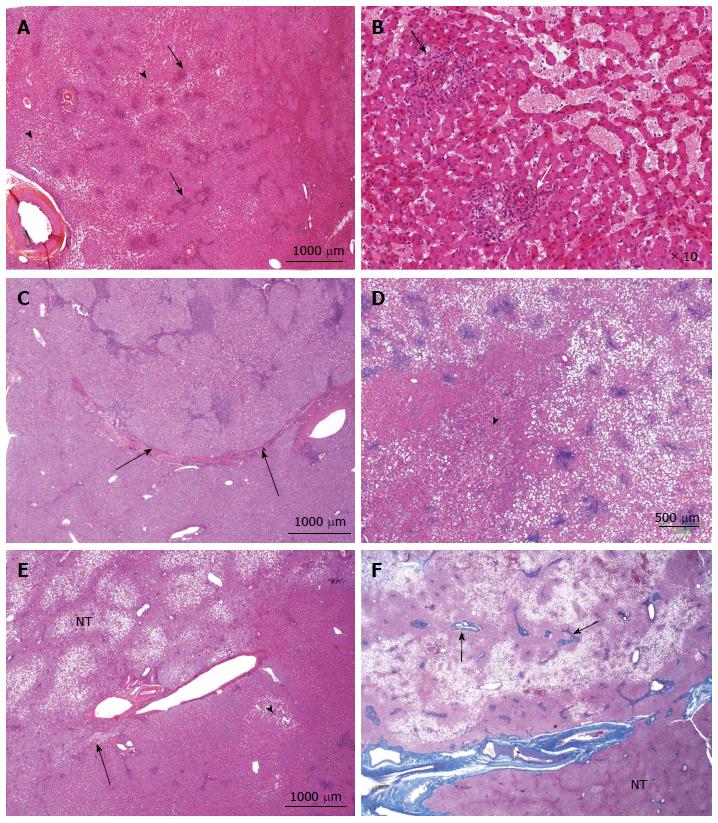
Figure 10 Inflammatory hepatocellular adenoma typical microscopic aspects.
A: Woman born in 1959; adenomatosis discovered by chance. Size of the largest nodule: 11 cm. Oral contraceptives for 23 years; BMI 27.0 kg/m2. Left hepatectomy 2008. HE: typical aspect of an inflammatory hepatocellular adenoma (IHCA) with many small inflammatory foci (arrows) dispersed within the tumor, associated with areas of moderate sinusoidal dilation ( arrowhead). In this area, sinusoids are dilated, another hallmark of this subgroup. B: Woman born in 1969; abnormal liver function tests. One liver nodule, 12 cm. Oral contraceptives for 16 years; BMI 26.0 kg/m2. Right hepatectomy 2004. HE: areas of sinusoidal dilatation and pseudo portal tracts with thick walled arteries and inflammatory cells (arrows), hallmarks of this subgroup. The diagnosis was confirmed by immunohistochemistry. C: Man born in 1968; abnormal liver function tests. One nodule 12 cm. BMI 30.0 kg/m2. Right hepatectomy 2011. HE: prominent inflammatory foci dispersed in the tumor; thick vessels at the border of the HCA (arrow). The diagnosis was confirmed by immunohistochemistry. D: Woman born in 1966; liver nodule, 3.5 cm discovered by chance. No oral contraceptives, BMI 24.5 kg/m2. Right hepatectomy 2004. HE: inflammatory foci, areas of sinusoidal dilatation; in this area, tumoral hepatocytes are steatotic. E: Woman born in 1973; overweight. Adenomatosis discovered by chance. Largest nodule 7 cm. Biopsy HCA. Tumorectomy IV, VI, VII 2003. HE: ill defined benign hepatocellular tumor (arrow); limited areas of sinusoidal dilatation (arrowhead) predominating at the periphery of the tumor. The non tumoral liver is steatotic, a frequent finding in this group of patients. F: Woman born in 1966; abnormal liver function tests. Several liver nodules. Biopsy HCA; oral contraceptives for 10 years, BMI 29.6 kg/m2. Left hepatectomy and tumorectomy IV and VI, 2007. Masson’s trichrome: pseudo portal tracts (arrows) with arteries in fibrous tissue; large areas of steatosis, within the tumor. The tumor is limited by thick arteries and veins from the non tumoral liver (NT).
- Citation: Sempoux C, Balabaud C, Bioulac-Sage P. Pictures of focal nodular hyperplasia and hepatocellular adenomas. World J Hepatol 2014; 6(8): 580-595
- URL: https://www.wjgnet.com/1948-5182/full/v6/i8/580.htm
- DOI: https://dx.doi.org/10.4254/wjh.v6.i8.580


