Copyright
©2014 Baishideng Publishing Group Inc.
World J Hepatol. Aug 27, 2014; 6(8): 580-595
Published online Aug 27, 2014. doi: 10.4254/wjh.v6.i8.580
Published online Aug 27, 2014. doi: 10.4254/wjh.v6.i8.580
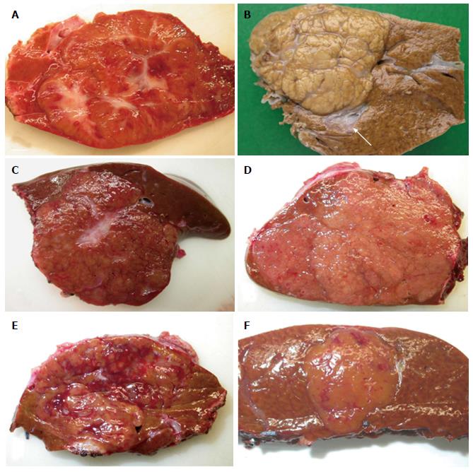
Figure 1 Focal nodular hyperplasia the macroscopic aspects.
A: Man born in 1988; 100 kg/1.95 m; abdominal pain; abnormal liver function tests; magnetic resonance imaging (MRI): liver mass 9.5 cm: Focal nodular hyperplasia (FNH)/hepatocellular adenoma (HCA) (bisegmentectomy in 2009). Fresh specimen: typical aspect of FNH: Tan, vaguely plurinodular tumor, non encapsulated, with central stellate scar. This macroscopic aspect is typical and the diagnosis of FNH is evident. The diagnosis was confirmed on HE and CK7. B: Woman born in 1951; acute abdominal pain followed by discomfort and pain on abdominal palpation; ultrasound (US): 2 nodules, largest one 3.5 cm. Magnetic resonance imaging (MRI): FNH (left hepatectomy in 2008). Fixed specimen: plurinodular tumor with thin fibrous bands, non encapsulated but well demarcated from surrounding liver parenchyma, with large portal tract at the interface (arrow). The diagnosis of FNH is evident. The diagnosis was confirmed on HE and CK7. C: Woman born in 1986; abdominal pain; MRI 2007: FNH 6.4 cm close to the biliary convergence; US in 2000: hemangioma 15 mm; 2004: 4.5 cm (tumorectomy in 2007). Fresh specimen: pedunculated irregular nodule with eccentric fibrous scar, well demarcated from the surrounding liver. The diagnosis of FNH is likely. D: Woman born in 1965; oral contraceptives for 18 years; abdominal pain; imaging (6 cm): FNH [surgery in 2005 (tumorectomy)]. Fresh specimen: clear-tan, vaguely plurinodular tumor, without clear-cut fibrous scar. The diagnosis of FNH is not self-evident. The diagnosis was confirmed on HE, CK7 and glutamine synthase (GS). E: Woman born in 1961; abnormal liver function tests; liver US: nodule 5.5 cm, MRI and US favors HCA over FNH (segmentectomy VII in 2009). Fresh specimen: irregular cut surface with tan nodules separated by congestive/reddish, atrophic areas. The diagnosis of FNH is unlikely. F: Woman born in 1956; check-up for arterial hypertension; liver imaging (2 cm nodule): probable HCA (tumorectomy in 2003) (other nodules were found). Fresh specimen: well-limited, non encapsulated, smooth nodule, with small reddish areas, without any fibrous bands or scar visible. The diagnosis of FNH is unlikely. The diagnosis was confirmed by HE, CK7 and GS.
- Citation: Sempoux C, Balabaud C, Bioulac-Sage P. Pictures of focal nodular hyperplasia and hepatocellular adenomas. World J Hepatol 2014; 6(8): 580-595
- URL: https://www.wjgnet.com/1948-5182/full/v6/i8/580.htm
- DOI: https://dx.doi.org/10.4254/wjh.v6.i8.580


