Copyright
©2014 Baishideng Publishing Group Co.
World J Hepatol. Mar 27, 2014; 6(3): 155-159
Published online Mar 27, 2014. doi: 10.4254/wjh.v6.i3.155
Published online Mar 27, 2014. doi: 10.4254/wjh.v6.i3.155
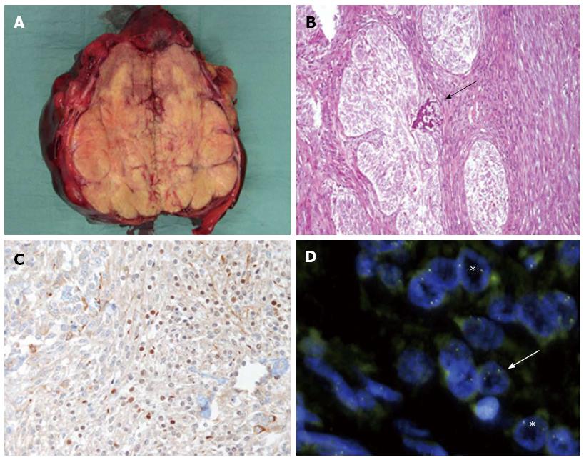
Figure 2 Pathological features of the reported calcifying nested stromal-epithelial tumor of the liver.
A: At gross inspection the lesion shows well defined margins and is arranged in lobules; B: At histology the lesion is characterized by the presence of well defined nests of epithelial cells separated by strand of stromal cells, focally admixed with calcifying deposits (arrow); C: Scattered neoplastic cells stain positive for WT-1 immunostaining; D: Some neoplastic nuclei showed well separated green and orange probes signals (arrow), in keeping with a translocation involving EWSR1 gene (FISH); some neoplastic nuclei (asterisks) did not show the same break apart pattern.
- Citation: Procopio F, Tommaso LD, Armenia S, Quagliuolo V, Roncalli M, Torzilli G. Nested stromal-epithelial tumour of the liver: An unusual liver entity. World J Hepatol 2014; 6(3): 155-159
- URL: https://www.wjgnet.com/1948-5182/full/v6/i3/155.htm
- DOI: https://dx.doi.org/10.4254/wjh.v6.i3.155


