
















©2013 Baishideng Publishing Group Co.
World J Hepatol. Aug 27, 2013; 5(8): 417-424
Published online Aug 27, 2013. doi: 10.4254/wjh.v5.i8.417
Published online Aug 27, 2013. doi: 10.4254/wjh.v5.i8.417
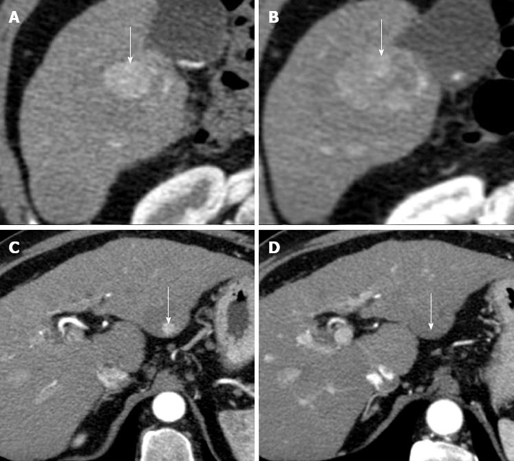
Figure 11 Hepatocellular carcinomas after sorafenib.
A: Pretreatment arterial phase computed tomography (CT) shows a large hypervascular hepatocellular carcinoma (HCC) (arrow) in right hepatic lobe; B: Arterial phase CT obtained at the same level of A, 9 mo after the start of sorafenib, shows increase in size of HCC (arrow); C: Pretreatment arterial phase CT image shows a second, small hypervascular HCC (arrow) arising within a larger hypoattenuating dysplastic nodule in left hepatic lobe; D: Arterial phase CT obtained at the same level of C, 9 mo after the start of sorafenib, shows disappearance of HCC enhancement (arrow). This case shows that different HCC response to sorafenib may occur in the same patient.
- Citation: Agnello F, Salvaggio G, Cabibbo G, Maida M, Lagalla R, Midiri M, Brancatelli G. Imaging appearance of treated hepatocellular carcinoma. World J Hepatol 2013; 5(8): 417-424
- URL: https://www.wjgnet.com/1948-5182/full/v5/i8/417.htm
- DOI: https://dx.doi.org/10.4254/wjh.v5.i8.417